Calcium

Dr Mc Blog


May 3, 2016
It was a very odd week. Monday started out with an African Grey parrot that was rushed in because of odd seizures observed by the owner. Popeye made the trip in a cat carrier since he couldn’t perch, and when I opened the grated door I had to laugh : a sizeable grey parrot with a dark maroon tail was fluttering convulsively but at the same time reaching out greedily to gobble down some peanuts just out of reach. He had figured out how to roll just right with each shake of his wings and was inching ever closer. Popeye wasn’t at all perturbed by his mild convulsions but was very upset that the peanuts kept evading him. After a brief discussion where I learned that he was fed almost exclusively on sunflower seeds I drew some blood tests and treated him for a calcium deficiency. Seeds may be high in protein and fat but are notoriously deficient in calcium and trace minerals. No single food item will ever provide complete nutrition to any animal. Popeye’s recovery was dramatic.

On Tuesday an owner presented his middle-aged Dachshund “Dracula” for a second opinion. The little dog just seemed to have muscle twitches all the time. He’d had some blood tests run, but nothing conclusive had turned up and regular anti-seizure medication had been of no help. Dracula lived a quiet life, mostly indoors and had never had any other medical issues. He trotted around the exam room, with a fine tremor marring his step. He wasn’t shaking from anxiety, nor did he act oddly in any other way. Looking over the chart shared by my colleague I noted that his blood calcium test was quite low. Since any single test may occasionally be off due to lab error I recommended we repeat these tests as a starting place. Again, his calcium levels came back very low. Diagnosis : A disorder of his parathyroid gland, which regulates calcium in the body. Treatment with calcium and Vitamin D supplements provided immediate relief while his case was fully worked up.

When the Green Iguana showed up on Wednesday I had a sneaking suspicion that I was having a “run” on calcium related issues. The lizards’ lower jaw was puffed out, looking mismatched to her upper face. She had an odd crook in her front leg from a “folding fracture”, indicating soft bones. Once again I investigated her diet and light exposure – never outdoors in natural sunlight (no creature was bred to live totally indoors!) and fed iceberg lettuce, no dark leafy greens, no vitamin supplements. Diagnosis : Metabolic Bone Disease or “Iguana Rickets”. Real sunshine and an improved diet did wonders for her.

By Thursday the staff had started to joke about it. Our 10 o’clock appointment was a young man with a muscular Pit Bull puppy, only 12 weeks old. The puppy had broken both his front legs. As I observed the heavy bodied male, named “Oscar”, I noted that he was unusually cheerful as he ran around the room, clumsily trying to chew on my assistant’s pants leg. He looked bow legged in front and indeed, both of his wrists (carpi) bulged and kind of knuckled over. He was walking on these legs, which a dog with a fracture usually cannot do, as it’s too painful and the bone actually can’t support weight as it slides apart. Questioning brought out the history of a puppy fed an exclusive diet of steak and hamburger. Oscar’s owner was proud of his beautiful dog and wanted to provide only the best for him. He had spared no expense by feeding an all-meat diet. What I was looking at was a very old-fashioned case of rickets or osteomalacia (soft bones). Without a balanced diet – and without bones meat alone is deficient in calcium – Oscar’s rapidly growing bones had softened and he couldn’t support the weight of his own body. The bones had started to fold down. Caught early, a corrected diet of simple commercial puppy food could reverse the process and over time the bones will straighten, though not always 100%. Happily, within two weeks Oscar was moving more normally.

The body is a marvel. Calcium is rich in many foods: dairy products like cheese, yogurt and milk, dark leafy greens, fish canned with their bones, bone meal by-products. Calcium is added to all pet foods formulated commercially, just as it is added to products like orange juice for people. But eating calcium rich food is not enough. The body requires Vitamin D3 to absorb and use the calcium taken in. Dogs, cats, birds and reptiles can all make their own Vitamin D when exposed to natural sunlight, through a complex interaction with UV light on their skin. Vitamin D is found naturally in egg yolk, fish oils and fatty fish like salmon or tuna, and cheese. Many animals, especially birds and reptiles, don’t naturally eat these foods and if kept deprived of sunlight (indoors/caged) will develop rickets, broken bones, and even seizures or death because they cannot make the Vitamin D they need. Even if their diet includes a calcium supplement they will suffer. For this reason special UV lights are recommended for indoor pets and at least weekly outings into real sunshine. All diets should include both fresh foods and commercial foods fortified with Vitamin D and Calcium, from turtle pellets to bird feed and other pet foods. Leave the balancing up to the professionals!

Christine B. McFadden, DVM
drmc@mcmenagerie.com


The Bladder

Dr Mc Blog


April 6, 2016
I thought we could talk about bladders today. Before you grit your teeth, let me say that it’s bound to be a lighter subject than body issues that lead to discussions of Milk of Magnesia or Preparation H. A far better organ system for public discussion. Bladders hold a deep fascination for my office staff. Instead of guessing how many jelly beans are in a jar, they often wait spell-bound to see how much urine that little dog can release on a waiting room plant. One client who frequents our hospital has a particularly clever little Yorkie who always manages to spray out a puddle despite our diligence and speed. Unfailingly, the good woman will say “My, he’s never done that before.”

Each day the kidneys filter the blood that flows through your veins, removing waste and impurities. Most of the blood continues to recycle to the rest of the body, but a small percentage of fluid and the filtered waste must be eliminated for continued good health. This fluid is called urine. In most mammals urine looks like a clear watery fluid, usually a shade of yellow, and is composed of excess body water, salts, and other waste. The yellow color is caused by the pigment urochrome, a breakdown product from hemoglobin. When dehydrated, the urine color may be darker yellow or orange as this pigment concentrates, indicating the need to drink more water. Each beat of the heart sends 20-25% of that blood flowing straight to the kidneys. Once urine is manufactured in the kidneys it travels from the kidneys via little tubes called ureters to the bladder, the holding tank in the body’s plumbing system. It is stored there until the pet voluntarily decides to empty their bladder, through another single tube called the urethra, to the outside. The act of emptying the bladder is called micturition or urination or voiding or peeing. Of course, all of these body parts can develop problems of their own, but the most common diseases revolve around the kidneys or the bladder. Kidney infections are not nearly as common as bladder infections, though often people confuse the two. Bladder infections may be caused by bacteria ascending up the urethra, because the bladder is the first stop from the “outside”. Today we will limit the discussion to dogs as the bladder problems seen in cats are often quite different in cause and treatment.

Prada’s Mom suspected a bladder infection when she observed blood in the urine. Female dogs are affected more often than males. Some affected pets may strain to urinate or may urinate very frequently or in unusual places. Although some dogs are depressed, Prada, a tiny snow-white Maltese, appeared bouncey and pain free. An examination was performed to determine if the bladder wall felt normal or was thickened. The bladder may have unusual “lumps” which could be cancerous growths or actual bladder “stones”, real rock-like growths that can fill the inside of the bladder. A urine sample was taken to analyze for bacteria, crystals, blood and other signs of disease. Sometimes the urine must be cultured to determine what kind of bacteria is affecting the dog and which antibiotic will best treat the infection. Because this was Prada’s second bladder infection in one year, a culture test was performed after obtaining her urine sample by cystocentesis, a sterile needle tap directly into the bladder. This method is preferred for a bacterial culture over a “catch” of urine because there will be no contamination from the pet’s hair or the environment as the sample is obtained. Chronic or repeat bladder infections may lead to the formation of bladder stones due to the chronic irritation of the bladder lining. These will require surgical removal in most cases. Bladder infections should never be minimalized as they can in turn ascend further up to the kidneys, which is potentially life threatening. Prada’s culture highlighted the best antibiotic choice for her and she responded quickly to her medication.

There is a small but significant obstacle to the pursuit and study of diseases of the bladder : obtaining the sample. Home catch, free catch, cystocentesis…. Apparently this has confounded veterinarians long before me! One very old veterinary text book neatly summed it up thusly : “Take about a dessert-spoonful of the dog’s urine (which you must use your own judgement how to obtain: place, for instance, a clean damp sponge at a spot where you think the animal will stale)…..”. Good to know I’m not alone!

Christine B. McFadden, DVM
drmc@mcmenagerie.com


Skin Care plus MRSA

Dr Mc Blog


March 1, 2016
Have you heard? There are new scrubs, mousses, lotions, creams, and wipes to improve your PETS’ appearance! For some reason these are not appearing in “Vogue” magazine or even “Dog Fancy”. Since I can’t skim a popular magazine in line at the grocery store without encountering pressure to rejuvenate my own skin, I don’t understand why all these products for pet appeal aren’t making big news. Perhaps skin care for animals just hasn’t caught on yet. Although I’m not entirely sure – I’ve heard there’s a blueberry facial mask out there, plus things like milk baths and rose water nail soaks for the (overly?) pampered pooch. Who knows? I heard of a doggie salon that gave the owners coupes of champagne while they waited. Perhaps that’s the force behind the trend.

For those of us in the less glitzy trenches, we refer to skin care as “dermatology” and the new mousses, wipes, etc have been formulated to address common health problems addressed daily in veterinary medicine.

Skin sores that bubble and break open on your pet may be the result of a bacterial staphylococcus infection. “Staph” is considered a normal inhabitant on the surface of the skin, until it goes rogue and breaks out as whiteheads or bubbling pus-filled lesions. In humans there is an increasing fear of an antibiotic resistant form of staph called “MRSA” (pronounce it “mersa”), which in simple terms means a staph bacteria that is resistant to the antibiotic methicillin, by extension the family of antibiotics headed up by penicillin. This bacteria takes advantage by invading the body through a wound, at a surgery site, or even a scraped elbow and is feared because it cannot be readily treated by the old tried-and-true antibiotics. The bacteria has simply found a way to survive, to overcome the tools fighting to destroy it. Because people in hospitals are at higher risk, due to their lowered immune resistance, stress, or fresh surgery sites, MRSA infections are feared. Pets can also develop a methicillin resistant staph infection, though it’s a slightly different bacterial strain called staph psuedointermedius, or MRSP (I say “mersip”. No one has corrected me yet). The veterinary community continues to monitor the prevalence of this infection and at this time it is not considered a risk to humans or other pets. When we find a skin infection not healing as expected we often recommend a culture swab be taken to diagnose both the type of bacteria and identify the best antibiotics to treat the problem. This is how we diagnose MRSP infections. One of the treatments is a mousse that cleans bacteria off the skin. It can also be used to bathe the pet, prior to an orthopedic or other major surgery, decreasing the risk of post-operative complications. There are anti-bacterial skin wipes, also.

Another new product is an eye wipe to assist dogs with epiphora, a condition where the pets’ tears spill down his or her face. A compound, porphyrin, in the tears causes a brown stain to form which is both unsightly and, staying wet, allows bacteria to breed which can lead to infection around the eye. The constantly wet skin may scald and create open sores under the eyes which is very painful. Dog and cat breeds with flat “pushed-in faces” like Pugs and Persians may be anatomically predisposed to problems of tears spilling out onto their faces. Once your veterinarian has determined that the tear ducts are open and draining properly and ruled out other medical causes, you may be directed to these wipes to keep the condition from escalating into painful sores.

Pets that are completely white haired, with no other color or pigment, are at higher risk of sun-related skin cancers as they sun bathe in our backyards here in the Central Valley. In cats red, scabby crusts may form on their ear tips or around the nose. In dogs it often begins on the thin skin of the nose or as deep purple blotches on the tummy near the back legs. Human sun screens may not be safe for your pets because they will eat it, literally, as they lick themselves! Zinc oxide (the white glob on the lifeguards’ nose) is toxic to dogs. Some dogs will tolerate hats or wear protective T-shirts, but the best idea in our hot summers is to simply keep these at-risk pets indoors during the day as much as possible.

In medicine there is always a new discovery around the corner. None of the skin care products I get to review will remove the laugh lines from Fido or Kitty but they do come with the promise of remedying a skin affliction. Please celebrate by raising your own glass of champagne.

Christine B. McFadden, DVM
drmc@mcmenagerie.com


Variety Is The Spice Of Life

Dr Mc Blog


February 17, 2016
They say variety is the spice of life. I am fortunate to have an extremely interesting career! In the past eight days I have seen one rabbit with an ear ache and spayed another. A lizard presented with scoliosis, curvature of the spine, caused by underlying malnutrition. I saw a cat with a very odd skin mite that may not have been diagnosed in Merced before! By the simple act of pressing scotch tape over the cat’s dander and looking at this under the microscope I found a mite I’d never seen before. My colleague, Dr. Klingborg, identified it from a derm (skin) meeting he’d attended in November and now we’ve held several discussions on the presence of this mite in Merced (it is considered to be contagious between cats). And always, the “usual” mix of cats, dogs, and birds. Today while performing spay surgeries (an ovariohysterectomy surgery) on cats I opened up one female cat only to find parts of a mummified fetus in her abdomen, causing no known distress to her before today. I completed the surgery and explained the situation to her owner, using digital photos taken during the surgery. Another veterinarian in the practice, Dr. Lauppe, was doing an orthopedic surgery on an injured knee while Dr. K removed a cancerous mass and attended to a dog that had been attacked by another dog (BBBD – Bit By Big Dog). A guinea pig with an eye ulcer came in and the Zoo needs a review on the Mountain Lion’s diet. A delightful Labrador had five enormous rotting teeth extracted and went home on antibiotics and painkillers. In two weeks he’ll be soooo much better! A lady called about a duck she thought might have eaten a tiny battery (the battery could corrode the stomach and cause heavy metal poisoning. All waterfowl are at risk of ingesting odd metal objects – they just like shiny objects). A woman (I never got her name) brought in a hummingbird that “fell from the sky”. We mixed up sugar water, revived the uninjured bird and sent it off with her to be released where she found it. An abuse case was brought in by Animal Control and a pregnant dog with three puppies stuck inside presented for a Caesarean section, performed beautifully by one of the other vets. I saw a Sulcata African tortoise with a mild upper respiratory disease and discussed the special dietary needs the new owner would want to address. Obtained from a Tortoise rescue group, the Sulcata’s shell mounded up on top, called pyramiding of the carapace. Much like the lizard with the deformed spine, it indicated an improper calcium and vitamin D ratio, the tip of the iceberg for signs indicating malnutrition.

February is National Dental Month for Dogs and Cats and most veterinarians who care for companion animals focus special attention on dental care this month. In veterinary medicine we are not focused on the whitest smile to impress someone! You are most fortunate if you have never experienced a tooth ache. Some dogs present with teeth so brown with mineralized plaque and calculus that it looks like moss has grown on them. That bad breath smell is from the bacteria growing along and under the gum line. The same bacteria that can dislodge and spread to the heart or kidneys. Cats demonstrate “jaw chatter” if you touch a tooth with a neck lesion cavity near the gum line – you can observe this incredibly painful response after you peel your cat off the ceiling. Most dogs and cats continue to eat despite foul teeth – please do not judge your pet’s dental pain by whether they haven’t starved to death! Over the years I have heard countless times of pets that “acted years younger” once their abscessed teeth were removed, their mouths healthy again. Call your vet today – there are some great deals out there this month!

Christine B. McFadden, DVM
drmc@mcmenagerie.com

Feline Urinary Syndrome or FUS, FIC, FLUTD

Dr Mc Blog


December 21, 2015
I think he was already 90 when we first met. The blue eyes, the felt hat reminded me of my own grandfather. A little shorter than me, he was a kind man who had moved to the States when his family emigrated from the Azores. It had been a long time since he worked on a dairy, and his companion now was a black and white cat, Jimmy, vividly marked like a Holstein cow. On that first meeting, Jimmy was just in for a check-up and vaccinations. We talked more about Mr. B’s wife, who had passed away after eating watermelon. He blamed himself for letting her eat the seeds – she loved watermelon so much that she’d inhale chunks heedless of any seed that went with it. He knew the seeds had killed her. I suspected cardiac arrest was the underlying cause from the rest of our discussion. That afternoon we forged a bond that grew over the years. Jimmy’s owner lived independently in a small house in the country. When he could no longer drive a car he rode a three wheeled bicycle, humming along the back roads for miles at a time.

Jimmy came and went as he pleased, growing a little plump under the tender loving care of home, plied with a variety of foods on a near daily basis. One morning Mr.B brought him in, both quite agitated. Jimmy was having trouble walking, crying out, and Mr. B said he kept trying to pee at home. He didn’t think anything was coming out.

When I placed my hand over the cat’s abdomen I immediately felt a hard ball inside, about the size of a baseball. Just that gentle touch yielded a scream of anguish from Jimmy that made us all jump. That ball was the cat’s bladder, swollen near to bursting because the outlet was obstructed. The tube that carries urine from the bladder to the outside is called the urethra. Tiny bladder crystals or struvite, sometimes called “sand” had formed in Jimmy’s bladder. The crystals precipitated from a combination of minerals found in his food, often magnesium, ammonium, phospate. Thirty years ago we called this the Feline Urinary Syndrome. We blamed the diet – no one knew why, or which cats – and knew it could occur in both male or female cats. The crystals irritated the lining of the bladder and urethra, causing swelling, thickening, and pain when urinating. In male cats, the urethra is longer and narrows at the tip, so they can obstruct. If a big clump of “sand” passes at one time it acts like a cork, stopping up the bladder so no urine can pass. Of course, the kidneys are the last to know, so they stay on the job making urine, which fills up the bladder until it can’t get any bigger (or it ruptures). Everything comes to a screeching halt and the kidneys start to shut down (post-renal kidney failure). At this point the cat has a relatively short time to get help – under 24 hours – before it dies.

Treatment at the veterinary hospital is immediate; this is not a procedure that can be safely put off until lunch. Risks are high. The cat must be anesthetized and a urinary catheter threaded carefully backwards up the urethra (retrograde) using a sterile flush if necessary to break down the obstruction. The catheter is placed directly into the bladder and sewn in place, often for 1 or more days. An intravenous catheter is also placed to immediately flush the system and get the kidneys up and running. Blood tests tell the picture of the degree of kidney disease. The cat will have more blood tests before coming off IV’s to be certain that kidney function has been regained. Oddly, most of these cases are not infected with bacteria, though we use antibiotics to prevent a secondary infection once an indwelling bladder catheter is in place. Other names for this syndrome now include Feline Interstitial Cystitis and Feline Lower Urinary Tract Disease.

A change in diet may prevent recurrence of crystal formation. This requires a Prescription diet from your veterinarian. The food maintains the urine PH in a very narrow range that prevents formation of more crystals. In dire circumstances when a cat obstructs a second time, a surgery called a perineal urethrostomy (big words!) is life-saving. The surgery widens the urethra 3-4x more than before. The cat may still form crystals, but it shouldn’t be able to obstruct again.

Jimmy responded well to treatment and readily accepted his new diet. He died from other causes about 2 years later. His owner grieved. It was springtime, and Mr. B had his door open to let in the fresh breeze. He dozed off in his recliner and was startled upon awakening to find a black and white cat, boldly patterned, winding herself through his legs and around the coffee table. He knew it wasn’t Jimmy, but? He named her Susie. She had come to stay. Every time I saw them together we would marvel at how Jimmy must have sent her.

FUS=feline urinary syndrome;FIC=feline interstitial cystitis;FLUTD=feline lower urinary tract disorder. If you like words.

Christine B. McFadden, DVM
drmc@mcmenagerie.com


Anesthesia : Science or Art?

Dr Mc Blog


October 13, 2015
The body doesn’t always perform, live or die, as expected. The medicine that worked before in a given situation, doesn’t. Or wondrously, against the textbook prediction (prognosis) a full cure is effected when…. It shouldn’t have? Of all the internal medicine problems, the surgeries, the diseases and treatments that veterinarians must try to explain to their clients, it is possible that anesthesia is the most difficult subject to explain and often creates more anxiety than the surgical procedure accompanying it.

Anesthesia is the administration of a chemical substance by pill, injection, or as an inhaled gas that will induce a state most similar to heavy sleep, but at a level of decreased consciousness where pain is not felt. This allows surgery to take place. “A chance to cut is a chance to cure!” was a motto endorsed by my surgical professors. Most veterinarians learn very quickly that no matter how great a surgeon they become, it is as nothing if their patient does not wake up afterwards. Anesthesia is indeed a very important factor in the final outcome.

There is much that goes into the training. With todays’ computer applications, you can punch in a pet’s weight, age and species and the computer will print out a list of combinations and choices of anesthetic for that particular patient. Ahhh, but here is where the art of science comes in. Science, applied to a living creature, is not that cut and dried. Is the patient very tiny or young? She might not take deep, regular breaths under anesthesia and may require assisted ventilation. How to best smooth out the adrenaline rush of a frightened pet so he’ll transition easily into anesthetic sleep? Is the patient very thin or very fat? A heart murmur? Epileptic seizures? So many different considerations must be factored in to choose a correct anesthetic regimen.  Each patient’s case is reviewed individually. Your veterinarian will perform a pre-operative exam looking for signs of underlying disease that might complicate anesthesia. What must be tolerated in an emergency will be refused for an elective surgery : for example, a cat with a snotty nose will be refused a spay surgery.

Most pets receive a preanesthetic medication, called a “pre-med”. This is often a mild tranquilizer or sedative mixed with medication to dry up respiratory secretions. The pre-med serves multiple functions : it dries up fluids, keeping the nose and airway passages clear and it helps to calm and quell that adrenaline rush, as  pre-medicated patients actually need less anesthetic agent throughout their surgery. Pre-emptive pain medication may be given at this early stage, which has been proven to tamp down pain so much before surgery that pain never peaks as high after surgery.

Pre-meds are given about thirty minutes before surgery. The drowsy patient is more relaxed for the second phase, anesthetic induction. Induction anesthetic brings the pet rapidly into deep sleep, often via an intravenous injection. Small patients may induce themselves into sleep with the same anesthetic gas that will be used throughout surgery. If induction is not followed by maintenance with a gas anesthetic it may only last for 5 minutes! This initial state of unconsciousness allows placement of an endotracheal tube, a long flexible tube through the mouth into the windpipe, or trachea, which creates a wide-open passageway for air, oxygen, and the anesthetic gas to be delivered. From this point your pet keeps itself asleep by regularly breathing this anesthetic gas in and out. Monitoring equipment that follows your pet’s heart rate and other parameters are used while under anesthesia. A variety of anesthetic gases are used today in veterinary medicine and can be reversed by simply disconnecting from the machine. The majority of pets in a short procedure, under one hour, will awaken in a few minutes and be walking within 15 minutes!

There is much anxiety about anesthesia, the fear of going to sleep and never waking up, of an unexpected death. The reality is that there are few complications with anesthetics using the drugs and protocols available today. Surgery is recommended for the health benefit of your pet, to repair broken bones, remove cancers, etc. and these benefits far outweigh the risk from anesthetic complications for most pets. Many of our pastimes, whether driving a car or playing in sports, are more risky. I sincerely hope this gives you a foundation for understanding the art and science of anesthesia.

Christine B. McFadden, DVM
drmc@mcmenagerie.com


Egg Bound Bird

When I first fielded the call I wasn’t overly concerned, probably because the caller was about 80 miles away and somebody else’s patient. It’s easy to give telephone advice. I am, in truth, happy to share knowledge that might benefit another veterinarian’s patient as it is by similar sharing that I have been taught how to heal.  So I carefully shared my experiences in Avian Obstetrics and wished all a good outcome. Then I cheerfully went about my own day.

Sometimes that is not the end of the story. Many days later I found myself enmeshed in the case personally. It started out simply enough. A young family owned several parrots. One of them, a delightful little green-cheeked Conure was a clown and gentle enough to tolerate their young toddler. The bird was named Salsa, for her green and red feathers. I say “her” loosely, because most birds in the psittacine or parrot family groups cannot be sexed visually and require a DNA blood test to know whether you have a boy or girl. We used to surgically sex birds years ago, entering the abdomen only on the left side because most birds have one sole functioning ovary, on the left. Males have two testes, located in the same area in the abdomen, in front of the kidneys, tucked up under their protective spine.

So nobody knew if Salsa was a boy or a girl, which hadn’t been very important until the family left for vacation and Salsa became ill.  She began to sit in the corner of the cage bottom, very quietly. Her breathing was fine, and if disturbed she was a little snippy but hopped about brightly. The babysitter noticed that Salsa was straining, maybe trying to poop, and her abdomen seemed distended. A quick trip to her regular veterinarian confirmed that Salsa had an egg stuck inside! (This also confirmed she was a female! Smile).

Welcome to the world of birds and their babies! Remember, birds are not mammals. Any female bird can make an egg, fertilization is not required. Nothing hatches from these eggs. The eggs produced by chickens for grocery store sale I call “baking” eggs, the kind you cook with. The formation of an egg is fascinating. The left ovary is attached to an oviduct, similar to the uterine horn in a dog and cat. The ovum or egg looks like a miniature yolk as it is first released into the oviduct, where it travels to the shell gland or uterus to become coated in calcium carbonate as a hard shell. The whole process takes around 24 hours. Most parrots lay an egg every 2 to 3 days, until they have a full “clutch” which completes their nest. Certain external stimuli such as increased sunlight, warmer temperatures and more food may promote egg production. In a pet bird there is no denying that egg laying can be a risky business.

Sometimes the egg shell is not properly hard, termed a “soft-shelled” egg. This is a warning sign that the bird may be malnourished, particularly deficient in calcium and vitamin D. Birds that live exclusively indoors and on seed diets are at high risk of this type of malnutrition. Calcium is needed not only for strong bones and firm egg shells, but also to produce the muscle contractions that will allow the egg to be laid. First time eggs may be too large to pass easily. Once the egg is stuck, things can go bad very quickly. The egg may be so large to prevent the bird from breathing easily. It may squish the intestines and block passage of fecal matter. The egg may press against the sciatic nerve, causing permanent left leg paralysis. It may lose moisture and become stuck to the tissues of the oviduct, eventually starting an infection that leads to death. Egg binding is a serious medical emergency in birds.

Salsa didn’t have a mate, so we knew this was just an empty egg inside her. Because Salsa couldn’t pass the egg on her own, her veterinarian, very appropriately, anesthetized the little Conure and attempted oocentesis, (don’t you love new words? Pronounce it “oh-oh-sent-e-sis”). A needle is inserted into the egg to withdraw the contents, in an effort to collapse and allow passage of the now much smaller egg. Salsa’s egg collapsed but did not pass. However, she was bright and perky once the pressure in her abdomen was relieved so no one was worried. When the owners returned home Salsa was delighted to see them. Over the next few days they noted, however, that she became increasingly less active, until finally she was sitting at the bottom of her cage again. Which is when she was referred to me, 17 days after becoming egg bound. I started her on antibiotics,  gave her injections of Vitamin D and Calcium, and rehydrated her with fluids before taking her to surgery. I use a surgical laser instead of a “cold steel” scalpel for surgical incisions as there is minimal blood loss. The oviduct with the collapsed egg and a second egg behind it (!) was isolated and carefully removed. The ovary was left in place. Her little abdomen was flushed and sutured up and within 15 minutes Salsa was back in her incubator, recovering from her “spay” surgery. At her recheck two weeks later her family reported that Salsa was back to her old self, all fun and games. She weighed exactly 65 grams on that visit, smaller than a cockatiel. Her diet had been fortified with pellets, fresh fruits, vegetables and a calcium supplement. Salsa celebrated her recovery with a pistachio nut!

****Please note : The images and video used in this post are NOT the same patient being discussed in the ‘Egg Bound Bird’ blog post. In the video, the obstetrical problem was managed medically. This video is shown with permission from the gracious owner of another egg bound patient. The Sun conure in the video is roughly twice the size of the little conure in the story, which required surgical intervention, similar to a C-section and hysterectomy. Now you can have visual aids to go along with the blog!

WARNING THE VIDEO BELOW CONTAINS SENSITIVE MATERIAL THAT MAY BE TOO MUCH FOR SOME TO VIEW.

Christine B. McFadden, DVM
drmc@mcmenagerie.com

Seizure Disorders

Dr Mc Blog


September 9, 2015
There was a tremendous commotion at the front door. I looked up to see a tiny ball of fur writhing in circles in the open doorway as her owner struggled to balance the door and simultaneously pick up her pet. Just as I reached them, the dog suddenly stopped her flailing, stood up, shook her coat and trotted the rest of the way in. Her owner looked up at me and shrugged wearily. “Coco does this every time she comes in for a bath.”

Really? Her dog had a SEIZURE every time she was groomed? I found this incredible. Actually, I didn’t believe it. There were some gaps in my medical training which Coco was going to neatly plug. Oh, I knew a lot about seizure disorders. A seizure or convulsion is when the brain has an electrical misfire and the body loses control, most often recognized when a pet falls to the ground, its legs jerking uncontrollably. In humans this is called a grand mal seizure. Any kind of animal may suffer from a seizure, sometimes as simple as staring blankly at a wall, to chasing butterflies to a full-blown fit.

There are many causes for seizures. One big group that lumps many chemicals together is “poison”. Thirty years ago strychnine poisonings were still being seen here in Merced – leftover coyote bait? I don’t know. Most of those chemicals are long gone and illegal today. Some have been replaced by less toxic yet effective products. The snail bait metaldehyde that can lead to seizures and death in pets is slowly disappearing as an iron chelate compound takes its place.

Many pets which have their first seizure at home scare their family no end, but are up and running around like normal by the time they arrive – emergency! at the clinic. Why do we ask you to come in? Because we don’t know if your dog will stop seizing on its own, and when the seizure continues over 10 minutes or longer, called status epilepticus, your pet may be at risk of dying. Dogs in status epilepticus, for example, may need to be completely anesthetized to allow his or her muscles to unclench from their rigor, and may have to be kept heavily sedated for a prolonged period of time. This requires hospitalization, IV fluid support and intensive nursing care.

If your pet DOES have a seizure, try to time it (yes, it feels like an eternity!). Most last less than 2 minutes. Do NOT try to grab your pet’s head – it may bite you. Dogs do not swallow their tongue while seizing. Most pets recover quickly from a seizure. If not on emergency, have your pet seen by a veterinarian as soon as possible.

If the pet is not still seizing when brought to the hospital we will want to run some blood tests. There are a wide variety of organic diseases that may lead to a seizure. Oddly enough, low blood calcium is one. Calcium is not only needed for strong bones, but it is vital for every muscle contraction, including the beating of the heart. A mother dog with a large litter of pups or possibly a poor diet may present in tetany when milk production drains her body of calcium. Low blood sugar is a common problem in toy breeds. Electrolyte disturbances, the body’s salts, can lead to seizure like activity. The liver, the pancreas, the parathyroid glands, the brain! It all becomes very complicated. Many times when we find a problem on the blood tests we can treat the underlying disease specifically and the seizures will disappear. Sometimes, when the blood tests are normal and the seizures recurrent, we diagnose Epilepsy. If there is reason to be concerned about brain cancer a test by CT brain scan is available for pets at several centers in the Valley.

Epilepsy is still incompletely understood. It may be inherited or may occur secondary to severe head trauma, like being kicked by a cow. Often we diagnose “idiopathic” epilepsy meaning “I don’t know why”. Most epileptic cases are treated with similar medications, regardless of underlying cause. Anticonvulsant medications work through their sedative effect on the brain, calming the electrical impulses so they do not cross the excitement or seizure “threshold” that results in a “fit”.

While the onset of most seizures is never understood – so there is no preventing nor anticipating the next one – Coco set out to prove that yes, doggie drama queens do exist! We ran her blood work. Normal. We started her on anticonvulsants. She seizured “like always” at her next bath. We changed meds. Another seizure with the suds. Her owner swore that Coco never had a seizure at home. We never saw a seizure with vaccinations! Not over a blood draw! Not with the clippers! Something about running water and shampoo just pushed her over the edge. It was not worth medicating her for such infrequent events. Though possibly less stylish, Coco was home bathed after that and sternly warned to stay out of mud puddles!

Christine B. McFadden, DVM
drmc@mcmenagerie.com


Uric Acid Stones and Spotty the Dalmatian


Dr Mc Blog

August 12, 2015
Well if you don’t know that “Spotty” is the perfect name for a Dalmation dog then you’re not a kid. Clarity is not reserved to children, but I find they are less likely to obfuscate descriptive terms. And a Dalmatian dog, though born white, is soon covered with a myriad of black (or brown) spots. This breed has been beloved for centuries, serving as a watch dog, a fire station dog and starring in a famous Disney movie. Going back to their origins, they were selected for their beautiful coats and rugged nature to accompany coaches, travelling long distances as guards. This led to them serving horse-drawn fire wagons, as they could keep up with the horses, barking an alert to clear people out of the way. At the scene of the fire they both calmed the horses and guarded the wagon. One tough, sturdy breed of dog. Despite the movie (all puppies are cute), Dalmatians are a breed highly unsuitable to be penned up in a house with little to do. No action hero likes to be sidelined. But the Dalmation mania has subsided and these days we see dogs well suited to their homes. Some even belong to firefighters!

Spotty was a family pet. He had recently celebrated his fifth birthday. He looked forward to his daily bowl of kibble and walk in the park. But….something seemed a little off. Spotty passed on his kibble one night and at the park he seemed to strain to urinate. In fact, he’d walk a few paces and lift his leg, but nothing came out. By the next morning Spotty wasn’t walking very well. His hind legs seemed stiff and he was panting and anxious. Sometimes his stomach would heave and it was hard to tell if he was trying to vomit or urinate but nothing came out either end. Spotty seemed painful. Time to go to the vet.

In the hospital it was obvious Spotty was in distress from a distance. Panting and unwilling to walk, the reason was soon obvious. His abdomen was mildly distended and palpation identified a very large, firm bladder. It hurt to touch and Spotty was in no mood to be brave. We had a strong suspicion as to the cause of Spotty’s problem.

You see, the Dalmatian breed of dog has a unique inherited predisposition to uric acid bladder stones. Foods like organ meat or seafood are high in a form of protein called purines. The liver is supposed to break this protein down into a harmless substance that the body absorbs but in Dalmatians, lacking this gene, the liver only processes the protein into uric acid, which is filtered through the kidneys and settles out as small stones (as hard as coarse sand) into the bladder. The best way I can draw this picture with words is to compare the Dalmatian, a mammal, to birds. Birds are uricotelic, meaning they do not break down body waste into urine but instead their kidneys excrete uric acid, the solid white matter you see in a bird dropping. This is normal for a bird. But mammals – dogs – are supposed to excrete waste filtered through the kidneys as a thin fluid, urine. Dalmatian dogs make urine, but some will also make uric acid crystals which build up in the kidneys and bladder. Hundreds of these crystals can be deposited inside the bladder and obstruct the male Dalmatian dog’s ability to urinate. Once the bladder outlet is obstructed the dog can’t urinate and the bladder will slowly fill as the kidneys continue to produce both urine and uric acid. Without relief of the obstruction the bladder will rupture and/or the kidneys shut down, both potentially leading to death.

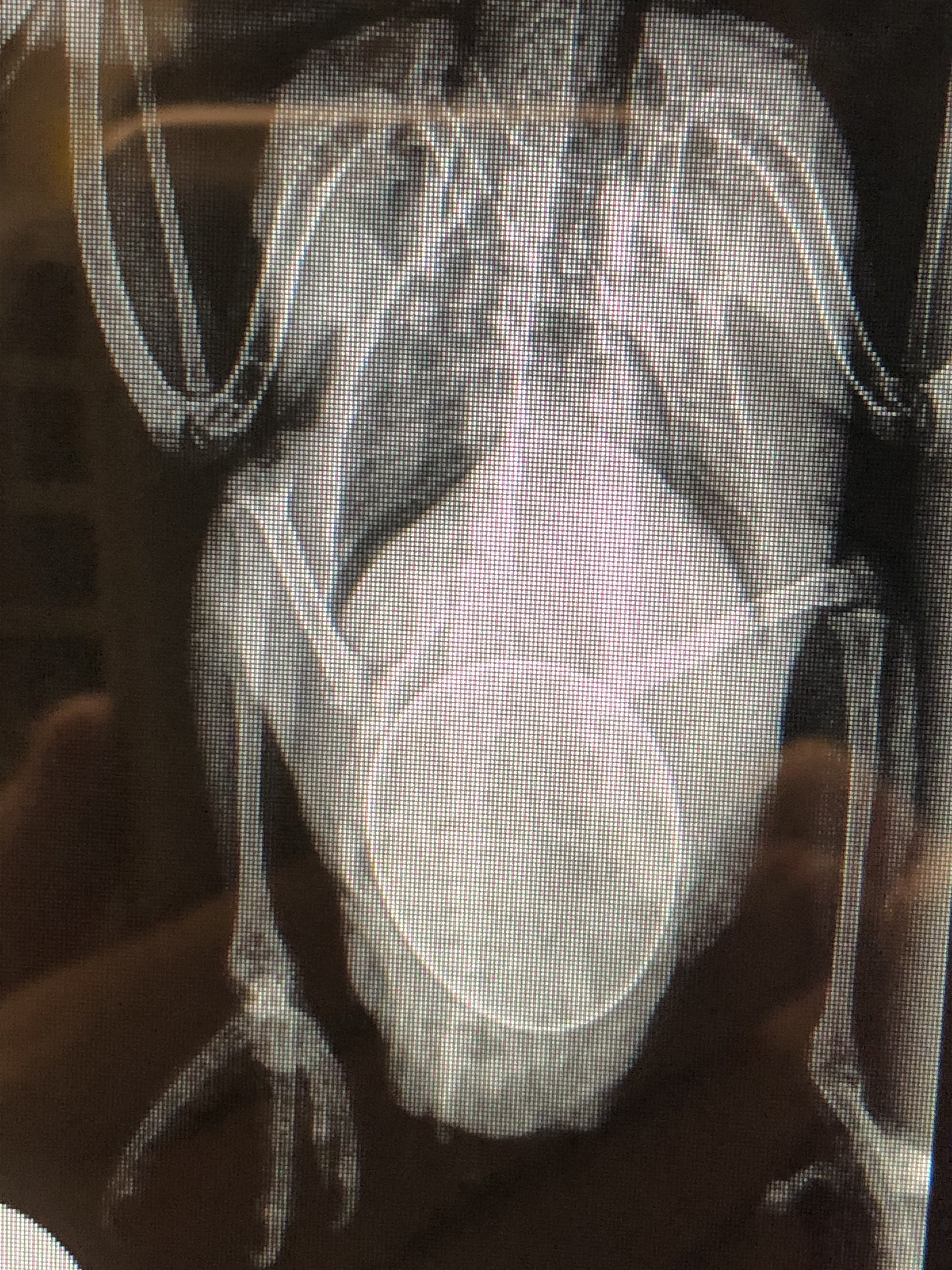
Spotty’s abdominal x-ray showed an enormous bladder, though the uric acid stones causing the problem weren’t well defined. His bloodwork demonstrated kidney distress, but within parameters that we felt could be reversed to normal once his obstruction was surgically cleared. We whisked Spotty in for emergency surgery, relieving his obstruction and emptying his bladder of hundreds of tiny grey stones. Intravenous fluids helped get his kidneys up and running again, a process we call diuresis. The stones or uroliths were analyzed to be certain they were uric acid in nature and a proper prevention plan put in place.

We know that high protein, meat-based diets may not be the best choice for any Dalmatian at this time as many are formulated with high purine foods of organ meat or seafood. Avoiding these purine-rich foods puts less burden on the liver and can greatly decrease the formation of stones. Dietary avoidance of these foods is a cornerstone to prevention.

This condition is similar to the disease of gout in people. In people the uric acid crystals settle in the joints, causing inflammation and pain. Interestingly, the same medication may be used to treat both humans and canines.

Spotty is lucky. Through a low-protein, non-meat based diet, medication and monitoring of his urine he is expected to lead a full, normal life. Because the gene has been identified, Dalmatian breeders can now specifically test and work to breed out this genetic mutation. The Veterinary Genetics Lab at the University of California, Davis, School of Veterinary Medicine offers genetic testing for a variety of breed-inherited diseases.

Christine B. McFadden, DVM
drmc@mcmenagerie.com


The Pancreas

Dr Mc Blog

July 28, 2015
Don’t you wonder how some things ever came to be named? Now the word “pancreas” comes from an anatomist named Ruphos of Ephesus, around 1 or 2 AD. In Greek pancreas means “all flesh”. And it IS a pale pink scrawny L-shaped ribbon of an organ sitting in a crook of small intestine near the right side of the stomach wall. Nothing you can really get your hands on during surgery. In fact, you don’t WANT to get your hands on it during surgery. The pancreas is notoriously sensitive to rough handling and maybe handling of any kind and as a young surgeon I worried that if I had to gently nudge it to one side to access some intestine or heaven forbid hooked it when trying to locate a recalcitrant uterine horn (uterus) during a spay surgery that my patient would have a belly ache for days afterwards or maybe die. That part turned out to be not so true but even after 30 some years of exploring abdomens I am still skittish about handling the pancreas and try to avoid any surgical contact.

So what does a pancreas do? The pancreas serves two very important yet distinctly separate functions in the body. One is an endocrine (hormonal) function, as the pancreas produces the hormone insulin. When this system runs amok, pets suffer from diabetes mellitus, often called “sugar diabetes”. The other cells of the pancreas exert exocrine (enzymatic) control and produce amylase and lipase, two enzymes that help digest food in the small intestine. When these exocrine cells malfunction, the digestive enzymes are released willy nilly into the pancreas, not the intestine, where they start to digest the wrong thing. This is the disease Pancreatitis. Inflammation of the pancreas and surrounding abdominal tissue as these enzymes eat away at it is so marked that it may be seen on an x-ray. Tremendous pain over the anterior abdomen is often present. The pancreas is most stimulated to produce its digestive enzymes by high fat foods and protein.

Kahlua was a doggie princess. A little pudgy in middle age, she was lovingly treated to her fair share of dog food and table goodies, often high in fat. One day she started to vomit her dinner. She was sluggish and screamed in pain when her owner gently picked her up around her tummy. In our office she was running a low grade fever and cried when her anterior abdomen was palpated. Among her blood tests a special test for pancreatitis, the Spec Cpl (specific Canine pancreatic lipase test) was positive. Both amylase and lipase scores were in the thousands. When we told her owner the news he was ghastly quiet. Finally he raised his eyes and told me quietly that he had just lost his wife to pancreatic cancer. Kahlua was all he had left of her. He didn’t need to say more. I couldn’t rule out cancer that early because there is no blood test to rule out cancer of the pancreas. If cancer is present, it may be the size of a small pimple and won’t always show up on an ultrasound scan. We would have to proceed with standard treatment for pancreatitis and if Kahlua did not improve as expected would review our other choices. After several days on intravenous fluids to flush out her body and absolutely no food (do NOT want to stimulate the pancreas during this time!) Kahlua brightened up considerably and was no longer painful over her tummy. Her signs of infection responded well to antibiotics and when we gave the all clear and offered a tiny meatball of a bland low fat food she gobbled it down and never looked back. Despite this brush with serious disease Kahlua never had another problem with her pancreas, though she did maintain a low fat diet for the rest of her long life. She was another lesson in proceeding cautiously when discussing any particular ailment of a pet that humans might share, as all too often I find that my diabetic patient has a diabetic owner, or that heart disease or cancer “runs in the family”. Kahlua was 7 years old when she developed pancreatitis and lived another 10 years beyond that, a princess to the end.

Christine B. McFadden, DVM
drmc@mcmenagerie.com