New Life for an Old Dog

She had been all black in her youth, but now at the age of 14 she is showing some gray around her muzzle. She used to jump and romp and play. Now she walks sedately across the yard, a faint weakness in her hindquarters. But always, Bella has class. Always, the gentle tail wag to let you know that she’s listening, she’s here.

Two years ago, Bella exhibited marked signs of difficulty when she tried to stand up in the morning. Upon examination, it was obvious that she was losing feeling in her back feet. Our X-rays showed degeneration of the spine in her back. We discussed options and Bella’s owners decided to see a specialist, where Belle underwent laminectomy spinal surgery. She recovered beautifully and even though she needed to stay on arthritis medications she moved quite fluidly again. Bella enjoyed her people and her life.

Bella is a Labrador Retriever, and though I don’t believe she has ever retrieved anything more interesting than a goodie from her owners’ hands, she shares a fate common to many Labs. Bred to be high energy dogs, capable of hunting all day and swimming in frigid waters to retrieve ducks, these dogs seem to naturally have an extra layer of protective fat. Given snacks and a soft life in the suburbs, many a Lab will put on a few extra pounds.

As I surveyed Bella in the exam room on Tuesday, I felt a deep unease. By no means obese, Bella could have stood to “lose a few” but not like this – Bella had lost 10 pounds over a two month period of time. Nothing in her diet or routine had changed, though her owners noted that she frequently passed a soft stool. Given her weight loss, I recommended that we start with a blood panel for diagnostic tests. Her physical exam was unremarkable for her age and existing medical history.

When the lab tests returned, the “biggies” were all good. No sign of diabetes, liver and kidney function within normal limits. Her body proteins were low. Hmmm.

Bella’s body either wasn’t making proteins or it was losing protein. In this case, with further testing, we were able to verify that she wasn’t losing protein in her urine, and that her small intestine wasn’t absorbing nutrients properly (her cobalamin levels, or vitamin B12, was too low). Of course, we knew she had chronic diarrhea, so it was only a small step towards the diagnosis of a protein-losing enteropathy, or PLE. Plowing ahead, what was the underlying cause of THAT? We asked that Bella return for abdominal x-rays and ultrasound. While corroborating thickened bowel loops, we also found a mass on her spleen. We didn’t know if that mass might be cancerous or not, but we hadn’t found any other signs of cancer and dogs can get along without their spleens very nicely. If it wasn’t cancer but a hematoma on the spleen, there was a risk of it rupturing and Bella bleeding to death. After several talks, we scheduled Bella for surgery. She would undergo a splenectomy to remove the spleen and have her intestine biopsied to confirm the cause of her PLE.

This was a lot to ask of an old dog. Bella was in surgery for over an hour, requiring her to lay on her back, not a terrific position for a dog that had undergone major back surgery. Positioned in a foam support tray throughout, I still worried. Surgery was successful and her recovery from anesthesia rapid.

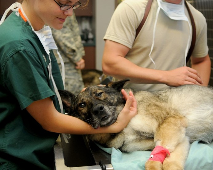
We kept her on intravenous pain meds, but there was no denying that Bella was slow to “bounce back”. Her owner cried that first day. Because of her IV fluids, Bella couldn’t move around easily. She struggled. We disconnected the fluids to take her outdoors to walk in the sunshine. Pain medication can cause a loss of appetite. I hand fed her. We slowly decreased the pain meds and upped her recreation. On Day four post-op, Bella met her owner in the parking lot. Moving slowly, she walked over to her, tail gently wagging. Bella went home. Her biopsy report diagnosed Inflammatory Bowel Disease (IBD) which responded well simply by feeding a specialty diet. The diarrhea disappeared. Her spleen was cancer free. Bella was on the road to a full recovery, as classy as ever.

Christine McFadden holds a license to practice veterinary medicine and surgery. She has cared for the family pets of Merced at Valley Animal Hospital for more than 30 years. Send questions or comments to drmc@mcmenagerie.com.
Cloning

For roughly $50,000 you can clone your favorite dog or cat. Speaking strictly for myself, I was somewhat startled. Are we there already?

Since I’m still sewing up cuts with plain old needle and thread, I find some of this high-tech stuff interesting. And amusing. And disturbing. So while I’m still healing the old fashioned way, somewhere out there veterinarians are offering to clone your pet.

They start with something as simple as a skin biopsy, a little 6 mm circle of tissue taken from your pet and carefully preserved for the genetic material found in its DNA. From this they can replicate the DNA and fuse the sample with an egg. This grows into an embryo that can be implanted into a surrogate mother. Once born you have a “clone” – a pet produced asexually from one ancestor to which they are genetically identical “twins.” A lot of this original work was being done in South Korea. There are now companies based in the United States that also offer either to preserve your pets’ DNA for future cloning or you can order up your very own clone today. In humans, identical twins result from the natural splitting of an egg in utero and both children contain identical DNA genetic makeup. Someone labeled identical twins “natural clones”?! Canine clones appear to have been the most popular pet clones requested by the public in this early work.

There’s a lot to consider here.

Say everything goes smoothly with the pregnancy and your little clone is born. Let’s name her “Clonia.” In the old nature-versus nurture-debate, right from the get-go we may have the same genetic material but it is going to be raised by another mother. That surrogate doggie mom may have been chosen for her availability as a uterus (think “animal shelter rescue”) and may not be particularly personable towards humans. And everyone, mom-dog and puppies, are living in a laboratory setting, not on Grandma’s comforter in your kids’ bedroom closet. So the first six weeks or so are not in a home environment and raise some serious questions about early socialization for the beloved Clonia. From what I could gather, responsible lab researchers provide careful monitoring to rule out abnormalities in the little clones, so many don’t go to their homes until they are several months old. Developmentally, they cannot be properly evaluated until then. This means even more time spent in a laboratory setting, coddled and cossetted, no doubt, for $50,000, but not a home. So when you do bring home your old pets’ genetic double, Clonia may not act exactly the same as her ancestor. Maybe not at all. Nurture versus nature.

And what happens to the mother dog, once she has performed her duties? This issue is clouded by lack of information. Clonia, by the way, cannot be registered with the American Kennel Club, no matter how prestigious her ancestral pedigree. Since the first dog cloned in the United States was born in February 2016, as of this writing, there isn’t a lot of information out there about “identical personalities” to the original pet. Somehow liking to chew up your sneakers or wagging your tail clockwise isn’t enough to convince me that you have reincarnated the original pet. Isn’t that really what this is all about? Clonia may not physically be an identical twin and again, her personality, shaped by very different early experiences, may be substantially different.

There are so many hearts and homes out there looking for each other that I find it hard to recommend seeking love through a clone. Would a clone of Shakespeare, father to twins Hamnet and Judith, still pen his famous works, such as “Romeo and Juliet” wherein he wrote “What’s in a name? that which we call a rose By any other name would smell as sweet.” I suspect that that which we call a dog is each an unique individual and that the search to duplicate your beloved pet by cloning borders on smoke-and-mirrors shell games, not something sweet.

Christine McFadden, DVM

drmc@mcmenagerie.com

Dog flu has been reported in the Valley. Here’s what you need to know.

Serious news for all dog owners in recent weeks, folks: Canine influenza, or dog flu, has been documented both in San Francisco and the Bay Area and in Fresno. Up to 50 cases were diagnosed in the Bay Area in mid-January, with a confirmed case in Fresno announced Jan. 24.

holding-dog-head-e1519278244598.jpg

This dog flu is highly contagious between dogs, with 80 percent of all exposed dogs becoming sick. Most of these dogs will be only mildly sick. Symptoms include coughing, sneezing, runny eyes and noses. Some dogs whose respiratory system has been attacked by the flu virus will develop secondary bacterial infections and have thick purulent (pus) discharge from their nose and a very high fever. Occasionally even pneumonia can occur and in rare instances, death.

You’ve all probably heard of “kennel cough,” which is a generic name for a variety of viruses or bacterial infections that cause a cough contagious to other dogs. The actual cause of bronchitis can only be determined through tests. An oral cheek and eye tear swab can be sent to the lab for a PCR test (polymerase chain reaction – if you really want to know more on that kind of testing, please research!) When last checked, a PCR upper respiratory test in dogs looks for up to 13 different viruses and other agents, including both forms of the Canine Influenza, H3N8 and H3N2. Dogs in close contact easily spread the germs by coughing on each other – in a kennel, a grooming parlor, at a dog park, day care or shelter. Anywhere dogs meet. In addition the flu virus can be spread by contaminated objects like food or water dishes, collars and leashes.

Should your dog contract canine influenza you will need to see your veterinarian. Treatment is tailored to symptoms and whether secondary infections are a concern. All affected animals should be isolated from other pets for 1 month to prevent the spread of this disease.

Canine influenza first made headlines in 2004 when several racing Greyhounds dropped dead in Florida. This was the CI H3N8. It wasn’t until March 2015 that there was another crisis when an epidemic broke out in dogs in Chicago. This was a different form of CI, H3N2. They thought part of the reason for an outbreak in a big city had to do with large concentrations of dogs moving through airports. The dog flu has not been widespread and risk to pets in many parts of the U.S. has been low.

The dog flu H3N2 has been known to affect cats. One outbreak occurred in cats in a high-stress shelter environment. No cats are known to have died from the canine influenza viruses, and there is no approved vaccine for cats.

Canine influenza viruses have not been found to be transmissible to humans.

Should you like more information on the canine influenza viruses please look up the American Veterinary Medical Association article on-line. Your local veterinarian has information and protective vaccine available to your pet. This bivalent vaccine protects against both strains of dog flu. Two vaccinations are required to start, given approximately one month apart, and can be started as young as seven or eight weeks of age. Because it takes a month for your pet’s body to mount a complete immune response, and given the closeness geographically of the epidemic, we recommend all dog owners contact their veterinarian to start protection. Be aware that like the Bordetella vaccine (a better known “kennel cough”) and human “flu shots” even with proper vaccination your pet might contract a mild case. Many area clinics are offering the vaccination to their regular patients. Simply put, we want to protect your pets. We’re here for you.

Christine B. McFadden

drmc@mcmenagerie.com

World Rabies Day


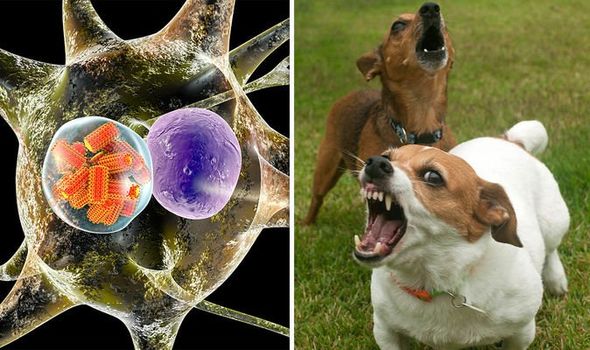
Several years ago I wrote an article on Rabies (Merced Sun Star, June 2014), a viral disease carried from animal to animal and sometimes from animal to human. For both animals and humans the disease is considered a death sentence.  I recently came upon a series of articles in a veterinary journal and was sad to learn that the disease is still a serious threat in much of the world and that approximately 60,000 people die from this disease every year. Still.


Why do I take up the story again? Because it deserves to be heard and, most importantly, it is still a threat to you and your loved ones. A very real threat if you enjoy traveling to out-of-the-way places.

Rabies-outbreak-map-rabies-symptoms-rabies-dog-bite-1125644

Rabies is a slow-moving viral disease. It can affect any mammal, but is usually spread from one carnivore to another as the virus is passed in saliva by bites through the skin. Infection after contact with infected saliva on an open wound is a possible, though unlikely, mode of transmission. Once a new victim has been bitten, the virus slowly makes its way into the nerves and through the nervous system to the brain. This can take two weeks to 2 months or longer. By the time the rabies virus is present in the saliva, death is near. It is for this reason that when an unknown dog bites a person the dog will be quarantined for 2 weeks.  Two weeks is long enough to monitor the dog : If rabid, the animal will die. The dog’s brain can then be tested for rabies virus and the bitten person still has ample time to receive the anti-rabies protective vaccine.


Rabies has been found in the wildlife in and around Merced County. It is endemic in area bats, raccoons, skunks and foxes. You, the pet owner, have set up a terrific protective line of defense by the simple stratagem of vaccinating your pet dogs and cats. It is believed that by vaccinating 80% of a populations’ pets an effective barrier is created to protect people against exposure to the disease. In the United States more rabid cats have been reported to the CDC (Center for Disease Control) than dogs for several years. Vaccinate your cats! Vaccinate all dogs and cats older than 3 months of age. Keep them up-to-date.


There are a few island communities which have managed to keep rabies out or eradicate its presence : Hawaii, Great Britain, and Japan are three. You cannot easily take a dog or cat into those Islands. I applaud their high security. The regions most likely to have rabid animals that endanger humans are underdeveloped countries in Asia and Africa.


For the traveler to a foreign country, be aware that the dogs roaming the streets may be high risk for encountering rabies. The unsuspecting traveler may admire a puppy, letting it lick their hands. Food you carry could attract attention from a roving dog. Many of these countries have inadequate medical services for diagnosing rabies or offering post-exposure prophylaxis (prevention/ PEP) services.  Rabies prevention is not universally available. The more exotic your outpost, the higher the risk of encountering rabies. Of the 60,000 people who die every year, nearly every single case (99%) of human death started with a bite from an infected dog.


Whether at home or traveling, camping for a weekend or following your dreams of adventure exploring a foreign land, please take precautions. Enjoy wildlife, but don’t treat or keep wildlife as pets. They cannot be vaccinated against rabies and may have variable incubation times for rabies. Avoid attracting wild life to your home or campsite. Do not feed wildlife. Do not feed your pets outdoors, as this attracts wild life and strays.  Avoid animals acting unnaturally. Animal-proof your trash. Avoid stray dogs and cats. Stray animals may not be vaccinated and they run high risk of exposure and contact with wild animals that might harbor rabies. The message, Backpackers : stay away from unknown dogs and wild life. The life you save might be your own!


World Rabies Day is Thursday, September 28th this year. This day is sponsored by an alliance among the World Health Organization and many other organizations working towards the goal of eradicating Rabies deaths in people and animals by the year 2030. They are joined by the One Health Initiative, a group dedicated to promoting increased communication and joint action between physicians and veterinarians globally through the integration of human medicine, veterinary medicine and environmental science. Rabies is cited as a classic example of what “One Health” is all about, as the control of rabies in animals has reduced the death toll in people from that disease. Together, we CAN make a difference.


Christine B. McFadden, DVM

drmc@mcmenagerie.com

Photo Credit : https://www.express.co.uk

The Large Animal Vet


Perhaps the title sounds misleading. A “large animal” vet develops a veterinary practice caring for the needs of livestock such as cattle (dairy or beef), sheep, goats and pigs. A “small animal” veterinarian like me is usually slotted into the care of companion pets such as dogs and cats. So when I examine a 200 pound dog, it is still a small animal and likewise there is often confusion about whom to call when the patient is a pot-belly/miniature pig. Not me.


The life of a small animal vet and that of a large animal practitioner are markedly different. One works almost exclusively indoors, dressed neatly, white coat in place. The other weathers life outdoors, facing extremes of blazing heat to icy cold, rain and snow. One always has a sink handy to tidy up. The other uses outdoor hoses more than they’d like. A dairy vet may check over 100 head of cattle in a morning and four “farm calls” make a full day. A small animal vet may follow the medical strands of more than 25 patients a day, winding through exams, blood results, x-rays and working in a surgery or two.  All juggle the demands of unexpected emergency work.


Being in an office most of the day, I rarely interact with my brother and sister large animal veterinarians. So when the pygmy goat from the petting zoo fell over dead I was unhappy, but proceeded to perform a post-mortem examination and sent tissue samples off to our regular lab. I had overseen the care of these goats for many years, but they were on the back burner when it came to my interest in the truly exotic Zoo collection. My concerns centered on whether or not the public might have fed something odd to the goat? (I’ll jump ahead here : No. The public did not harm the goat). I mulled over the problem but prepared to wait until the pathology report was finished sometime in the next week. And then another goat died.


I was unnerved. The goats were being closely watched and none had shown any outward signs of sickness. They had all lived for more than 8 years at the Zoo and had never, collectively, suffered a single injury amongst themselves. I immediately sought the expertise of a Large Animal veterinarian. The Goat Expert was on a dairy farm. The return call came in as I was finishing one surgery and about to start another. Gloves still on, I was staring at some x-rays

in-between these surgeries when they told me they had Dr. B on the line. Multi-tasking at its best.


I hurried over, snapping gloves into the waste receptacle and grabbing pen and paper. I introduced myself and LAUNCHED into a recital of my goat woes. I verified that he had goat experience? Yes. I gave him dates, genders, date of deaths, lack of lab results, still pending. I drew breath to spew forth another list of details and heard him gently respond, “Yes…. I think…. I might be able…. to help”. His measured tones were from a man used to the gentle rhythms of milking machines, contented cows swishing their tails, chewing their cuds. Da-dum da-dum to my staccato dop-dop-dop-dop-dop! I managed to squeeze in a few more hurried sentences (surgery! Waiting!) before he responded calmly, “I think I drive past your practice on my way up from this dairy….”. I opened my mouth. Closed my mouth. I could hear the clouds, feel the sunshine, almost see the shining black and white hides of the gentle Holsteins he surveyed as we spoke. It was all there in the rhythm of his speech. I told myself to stop yapping before he decided my goat problem would be too stressful for him to bother with.


An hour later Dr. Thomas Bauman drove up in a large truck outfitted for all manner of veterinary ministrations. He spent an hour and a half doing a post-mortem on one goat. He had a wicked knife and mulled over the cause of sand in the stomach. He felt it was too much. Did we feed on the ground? No. But little kids feed the Zoo goats oat hay pellets and they often dropped to the sand, with all the goats scrambling to get their share. Hmmm. He gave the problem his full attention and we submitted a gazillion samples to the State Lab, including an intact eyeball because it would be useful for trace metal analyses. The final answer was a copper deficiency in the feed, to which Pygmy goats are especially susceptible. Hay grown in the San Joaquin Valley is often deficient in copper. The salt lick fed at the Zoo did not have added copper because Alpacas are in with the goats and they are susceptible to copper toxicity (too much) if supplemented. So we now feed our goats little capsules of copper wire every six months and all is good. But sometimes I find myself wishing that I was a Large Animal veterinarian. Just so I could slow down and smell the …..never mind.


Christine B. McFadden, DVM

drmc@mcmenagerie.com

Accreditation / Monkey Pox Virus

Dr Mc Blog


September 14, 2016
I am going to share with you the Veterinary Accreditation Program through the United States Department of Agriculture (USDA) and the Animal and Plant Health Inspection Service (APHIS). What? The USDA-APHIS educate and certify veterinarians to inspect animals for disease prior to writing a health certificate for animal travel.

The Center for Disease Control (CDC) in Georgia is a governmental body we should all get down on our knees and give thanks for! The CDC is our nation’s health protection agency. It monitors all kinds of diseases that crop up in the United States and abroad that might affect us, from food borne illnesses (think E coli in the spinach or listeriosis in raw cheese products) to contagious measles outbreaks that start at California resorts to zoonotic infections passed from animals to people. The CDC works hand in hand with the USDA-APHIS, and the system trickles down to yours truly, the local town veterinarian. Which means I am authorized to fill out and sign a health certificate for a pet planning to travel to another state or country. Not a big deal? Think again.

Why so much interest you might well ask, in where Fido goes on vacation? Is this just another way that Big Brother is invading your privacy? The short answer is no. According to the USDA-APHIS they are recognizing new emerging infectious diseases world-wide. Of ALL the bacteria, viruses and other disease-causing pathogens that affect humans, they estimate 61% to be zoonotic or spread from animals to humans. Of the new “emerging” diseases they believe at least 75% to be zoonotic. The USDA-APHIS bodies wish to protect YOU by verifying that all animals that travel are disease-free, to safeguard humans. As a veterinarian may I add that this also helps safeguard our pets and our food sources. Our way of life.

The National Veterinary Accreditation Program web site shares some fascinating information of how the local town veterinarian can be a link in this chain of protection.

In 2003 a shipment of over 700 small mammals and rodents were imported to the US from Africa. The wildlife importer brought in dormice, various unusual rats and some rope squirrels. There is always a market for unusual animals in the pet trade, sometimes zoological parks or animal preservation parks, even research. The animals arrived safely in Texas, where they were re-distributed to Illinois. Many of the rats were housed near Prairie dogs. The Prairie dog has been adopted as a pet by many people. They are native to many areas of the United States. They have not been bred in captivity for generations, but may be relatively docile, eat timothy hay and are awake in the daytime which is a real bonus for people who enjoy interactive time with small rodents as pets. Unbeknownst to all, the rats were carrying the monkeypox virus and transmitted it to the Prairie dogs. The Prairie dogs were distributed to pet shops in six Midwestern states. These Prairie dogs went into pet homes.

Monkeypox is a virus in the same family as Smallpox. Smallpox was a deadly viral disease that the European explorers and settlers first brought to the Americas in the 1600’s. Native American tribes had never encountered this virus and had no natural immunity; many Indian tribes were decimated by Smallpox. Smallpox was eradicated in the United States before 1980, so a vaccination program against Smallpox is no longer enforced. Monkeypox was first diagnosed in Africa over 40 years ago. The disease causes a fever, enlarged lymph nodes and small raised polka-dot like spots or rash that may cover the body. Both humans and animals can develop the rash. Monkeypox is known to be zoonotic, spreading from a sick pet to a human by body fluids, soiled bedding and close contact. Signs of Monkeypox can show up as little as one day after exposure. There is no specific treatment against this virus, though vaccination against Smallpox may provide protection against Monkeypox for 85% of the people who receive it. Again, no one has routinely received Smallpox vaccination since 1972. Children born after that time will have no immunity.

Monkeypox had NEVER been seen or diagnosed outside of continental Africa until this outbreak in 2003 in the United States. Over a period of four weeks, from May to June, at least 47 people were infected from their new Prairie dog pets. One of the first family cases started May 11th 2003 when two Prairie dogs arrived in their new home. One of the Prairie dogs was ill with a skin rash and runny nose on the 15th and taken to their veterinarian. By the 20th the family was ill and the Prairie dog died. The veterinarian sent tissue samples to various labs at the same time as biopsies of the skin were being performed on the family by a dermatologist. Joint diagnosis confirmed Monkeypox. Quick action taken by the CDC, USDA and APHIS contained the outbreak. No person died. Monkeypox has not been seen in the United States since.

As a small town veterinarian I have been involved with potential cases of Rabies, Salmonella, Campylobacter, Avian tuberculosis, Chlamydiosis and West Nile disease. All veterinarians are here to serve.

Christine B. McFadden, DVM
drmc@mcmenagerie.com


Bupthalmia

Dr Mc Blog


August 16, 2016
Somewhere along the way your children catch on to you and become very suspicious when you are happy about something. Mine questioned the origin of names I kept pressing upon them for new pets. They obstinately refused to name the new cat “Tenesmus”. C’mon, I begged, it sounds great, doesn’t it? What does it mean? they questioned. Surly attitude! When I confessed tenesmus meant “straining to defecate” they did not laugh. I told them no one else would know. “No, Mom”, they said.

I have another favorite : Buphthalmia. Yes, it sounds like a long burp! Is there anyone else out there who only burps after drinking root beer? I digress. So what is “buphthalmia”? Pronounce it “bup” then add “thal-mee-ah”. It was an ophthalmologist, an eye doctor, who coined this word. Spelling that ph-th thing was the first question on our opth exam back in vet school.

Buphthalmia is different than exophthalmia. Exophthalmia is when the eyeball bulges out of the orbit, pushed behind by a space-occupying lesion like a blood clot or a mass. Buphthalmia is a little different : you will see an enlarged eyeball, bigger and wider than normal, because the cornea and surrounding tissues covering the eyeball have been stretched out by fluid build-up inside the eye.

The eyeball is nestled safely inside a protective ring of bones, often collectively referred to as the eye socket or orbit. The eye is an amazing organ and provides one of the five major senses, sight. The eyes of many animals have specialized features that allow them to see underwater, see in the dark, reflect light, and see in color. Most mammals and many birds and reptiles have a third eyelid which slides over their eyeball to both protect and lubricate the eye. Humans lack a third eyelid. It slides up from inside the inner lower corner of the eye, near the nose, and may be light pink in color. The inside of the eye is constantly bathed in a gentle fluid, the aqueous humor. This fluid circulates in front of the lens and helps to keep the eyeball round. The fluid pressure provided by the aqueous humor can be measured as intra-ocular pressure or IOP. If you, a human, has ever seen an eye doctor, they have probably measured your IOP with a tonometer – a little puff of air directed straight at the center of your cornea. We also use tonometry in veterinary medicine. Why do we care? Because the fluid in the front of the eyeball can build up, creating too much pressure. Too much pressure inside the eye is diagnosed as GLAUCOMA. This often happens because the aqueous humor doesn’t drain away properly. Once clogged, through a disease process or even an inherited problem of narrow drainage, the fluid builds up, creating pressure on the outer eyeball membranes and cornea which spread and bulge. The eye may look red or bluish and the pupils may dilate. Once the eyeball has enlarged, called buphthalmia, there is no going back. The eyeball will never return to its original size.

Your pet has an emergency. Without immediate medical treatment, in as little as a couple of hours the sustained high intraocular pressure can lead to blindness. If only one eye is affected by glaucoma the other may be at risk. Buphthalmia is a classic sign of glaucoma. Once diagnosed, the remaining Healthy eye should be treated with preventative eye drops for the rest of the pet’s life.

Certain breeds are predisposed to glaucoma, though the largest category is “mixed breed”, so no dog is safe. Cocker Spaniels, Bassets and Beagles, Boston Terriers and Norwegian Elkhounds all make the list. I’ve seen it in a family of Bouviers, an Akita and in Jack Russell Terriers. One day the owner notes their dog’s eye is a little cloudy and the next day as they come in for their appointment it is bulging and blood shot and ugly. It is too late to save the eye. A blind, painful eye requires enucleation or removal, for humane reasons. Thankfully, many eyes can be saved by prompt attention and treatment.

Christine B. McFadden, DVM
drmc@mcmenagerie.com


Skin Care plus MRSA

Dr Mc Blog


March 1, 2016
Have you heard? There are new scrubs, mousses, lotions, creams, and wipes to improve your PETS’ appearance! For some reason these are not appearing in “Vogue” magazine or even “Dog Fancy”. Since I can’t skim a popular magazine in line at the grocery store without encountering pressure to rejuvenate my own skin, I don’t understand why all these products for pet appeal aren’t making big news. Perhaps skin care for animals just hasn’t caught on yet. Although I’m not entirely sure – I’ve heard there’s a blueberry facial mask out there, plus things like milk baths and rose water nail soaks for the (overly?) pampered pooch. Who knows? I heard of a doggie salon that gave the owners coupes of champagne while they waited. Perhaps that’s the force behind the trend.

For those of us in the less glitzy trenches, we refer to skin care as “dermatology” and the new mousses, wipes, etc have been formulated to address common health problems addressed daily in veterinary medicine.

Skin sores that bubble and break open on your pet may be the result of a bacterial staphylococcus infection. “Staph” is considered a normal inhabitant on the surface of the skin, until it goes rogue and breaks out as whiteheads or bubbling pus-filled lesions. In humans there is an increasing fear of an antibiotic resistant form of staph called “MRSA” (pronounce it “mersa”), which in simple terms means a staph bacteria that is resistant to the antibiotic methicillin, by extension the family of antibiotics headed up by penicillin. This bacteria takes advantage by invading the body through a wound, at a surgery site, or even a scraped elbow and is feared because it cannot be readily treated by the old tried-and-true antibiotics. The bacteria has simply found a way to survive, to overcome the tools fighting to destroy it. Because people in hospitals are at higher risk, due to their lowered immune resistance, stress, or fresh surgery sites, MRSA infections are feared. Pets can also develop a methicillin resistant staph infection, though it’s a slightly different bacterial strain called staph psuedointermedius, or MRSP (I say “mersip”. No one has corrected me yet). The veterinary community continues to monitor the prevalence of this infection and at this time it is not considered a risk to humans or other pets. When we find a skin infection not healing as expected we often recommend a culture swab be taken to diagnose both the type of bacteria and identify the best antibiotics to treat the problem. This is how we diagnose MRSP infections. One of the treatments is a mousse that cleans bacteria off the skin. It can also be used to bathe the pet, prior to an orthopedic or other major surgery, decreasing the risk of post-operative complications. There are anti-bacterial skin wipes, also.

Another new product is an eye wipe to assist dogs with epiphora, a condition where the pets’ tears spill down his or her face. A compound, porphyrin, in the tears causes a brown stain to form which is both unsightly and, staying wet, allows bacteria to breed which can lead to infection around the eye. The constantly wet skin may scald and create open sores under the eyes which is very painful. Dog and cat breeds with flat “pushed-in faces” like Pugs and Persians may be anatomically predisposed to problems of tears spilling out onto their faces. Once your veterinarian has determined that the tear ducts are open and draining properly and ruled out other medical causes, you may be directed to these wipes to keep the condition from escalating into painful sores.

Pets that are completely white haired, with no other color or pigment, are at higher risk of sun-related skin cancers as they sun bathe in our backyards here in the Central Valley. In cats red, scabby crusts may form on their ear tips or around the nose. In dogs it often begins on the thin skin of the nose or as deep purple blotches on the tummy near the back legs. Human sun screens may not be safe for your pets because they will eat it, literally, as they lick themselves! Zinc oxide (the white glob on the lifeguards’ nose) is toxic to dogs. Some dogs will tolerate hats or wear protective T-shirts, but the best idea in our hot summers is to simply keep these at-risk pets indoors during the day as much as possible.

In medicine there is always a new discovery around the corner. None of the skin care products I get to review will remove the laugh lines from Fido or Kitty but they do come with the promise of remedying a skin affliction. Please celebrate by raising your own glass of champagne.

Christine B. McFadden, DVM
drmc@mcmenagerie.com


Variety Is The Spice Of Life

Dr Mc Blog


February 17, 2016
They say variety is the spice of life. I am fortunate to have an extremely interesting career! In the past eight days I have seen one rabbit with an ear ache and spayed another. A lizard presented with scoliosis, curvature of the spine, caused by underlying malnutrition. I saw a cat with a very odd skin mite that may not have been diagnosed in Merced before! By the simple act of pressing scotch tape over the cat’s dander and looking at this under the microscope I found a mite I’d never seen before. My colleague, Dr. Klingborg, identified it from a derm (skin) meeting he’d attended in November and now we’ve held several discussions on the presence of this mite in Merced (it is considered to be contagious between cats). And always, the “usual” mix of cats, dogs, and birds. Today while performing spay surgeries (an ovariohysterectomy surgery) on cats I opened up one female cat only to find parts of a mummified fetus in her abdomen, causing no known distress to her before today. I completed the surgery and explained the situation to her owner, using digital photos taken during the surgery. Another veterinarian in the practice, Dr. Lauppe, was doing an orthopedic surgery on an injured knee while Dr. K removed a cancerous mass and attended to a dog that had been attacked by another dog (BBBD – Bit By Big Dog). A guinea pig with an eye ulcer came in and the Zoo needs a review on the Mountain Lion’s diet. A delightful Labrador had five enormous rotting teeth extracted and went home on antibiotics and painkillers. In two weeks he’ll be soooo much better! A lady called about a duck she thought might have eaten a tiny battery (the battery could corrode the stomach and cause heavy metal poisoning. All waterfowl are at risk of ingesting odd metal objects – they just like shiny objects). A woman (I never got her name) brought in a hummingbird that “fell from the sky”. We mixed up sugar water, revived the uninjured bird and sent it off with her to be released where she found it. An abuse case was brought in by Animal Control and a pregnant dog with three puppies stuck inside presented for a Caesarean section, performed beautifully by one of the other vets. I saw a Sulcata African tortoise with a mild upper respiratory disease and discussed the special dietary needs the new owner would want to address. Obtained from a Tortoise rescue group, the Sulcata’s shell mounded up on top, called pyramiding of the carapace. Much like the lizard with the deformed spine, it indicated an improper calcium and vitamin D ratio, the tip of the iceberg for signs indicating malnutrition.

February is National Dental Month for Dogs and Cats and most veterinarians who care for companion animals focus special attention on dental care this month. In veterinary medicine we are not focused on the whitest smile to impress someone! You are most fortunate if you have never experienced a tooth ache. Some dogs present with teeth so brown with mineralized plaque and calculus that it looks like moss has grown on them. That bad breath smell is from the bacteria growing along and under the gum line. The same bacteria that can dislodge and spread to the heart or kidneys. Cats demonstrate “jaw chatter” if you touch a tooth with a neck lesion cavity near the gum line – you can observe this incredibly painful response after you peel your cat off the ceiling. Most dogs and cats continue to eat despite foul teeth – please do not judge your pet’s dental pain by whether they haven’t starved to death! Over the years I have heard countless times of pets that “acted years younger” once their abscessed teeth were removed, their mouths healthy again. Call your vet today – there are some great deals out there this month!

Christine B. McFadden, DVM
drmc@mcmenagerie.com

Feline Urinary Syndrome or FUS, FIC, FLUTD

Dr Mc Blog


December 21, 2015
I think he was already 90 when we first met. The blue eyes, the felt hat reminded me of my own grandfather. A little shorter than me, he was a kind man who had moved to the States when his family emigrated from the Azores. It had been a long time since he worked on a dairy, and his companion now was a black and white cat, Jimmy, vividly marked like a Holstein cow. On that first meeting, Jimmy was just in for a check-up and vaccinations. We talked more about Mr. B’s wife, who had passed away after eating watermelon. He blamed himself for letting her eat the seeds – she loved watermelon so much that she’d inhale chunks heedless of any seed that went with it. He knew the seeds had killed her. I suspected cardiac arrest was the underlying cause from the rest of our discussion. That afternoon we forged a bond that grew over the years. Jimmy’s owner lived independently in a small house in the country. When he could no longer drive a car he rode a three wheeled bicycle, humming along the back roads for miles at a time.

Jimmy came and went as he pleased, growing a little plump under the tender loving care of home, plied with a variety of foods on a near daily basis. One morning Mr.B brought him in, both quite agitated. Jimmy was having trouble walking, crying out, and Mr. B said he kept trying to pee at home. He didn’t think anything was coming out.

When I placed my hand over the cat’s abdomen I immediately felt a hard ball inside, about the size of a baseball. Just that gentle touch yielded a scream of anguish from Jimmy that made us all jump. That ball was the cat’s bladder, swollen near to bursting because the outlet was obstructed. The tube that carries urine from the bladder to the outside is called the urethra. Tiny bladder crystals or struvite, sometimes called “sand” had formed in Jimmy’s bladder. The crystals precipitated from a combination of minerals found in his food, often magnesium, ammonium, phospate. Thirty years ago we called this the Feline Urinary Syndrome. We blamed the diet – no one knew why, or which cats – and knew it could occur in both male or female cats. The crystals irritated the lining of the bladder and urethra, causing swelling, thickening, and pain when urinating. In male cats, the urethra is longer and narrows at the tip, so they can obstruct. If a big clump of “sand” passes at one time it acts like a cork, stopping up the bladder so no urine can pass. Of course, the kidneys are the last to know, so they stay on the job making urine, which fills up the bladder until it can’t get any bigger (or it ruptures). Everything comes to a screeching halt and the kidneys start to shut down (post-renal kidney failure). At this point the cat has a relatively short time to get help – under 24 hours – before it dies.

Treatment at the veterinary hospital is immediate; this is not a procedure that can be safely put off until lunch. Risks are high. The cat must be anesthetized and a urinary catheter threaded carefully backwards up the urethra (retrograde) using a sterile flush if necessary to break down the obstruction. The catheter is placed directly into the bladder and sewn in place, often for 1 or more days. An intravenous catheter is also placed to immediately flush the system and get the kidneys up and running. Blood tests tell the picture of the degree of kidney disease. The cat will have more blood tests before coming off IV’s to be certain that kidney function has been regained. Oddly, most of these cases are not infected with bacteria, though we use antibiotics to prevent a secondary infection once an indwelling bladder catheter is in place. Other names for this syndrome now include Feline Interstitial Cystitis and Feline Lower Urinary Tract Disease.

A change in diet may prevent recurrence of crystal formation. This requires a Prescription diet from your veterinarian. The food maintains the urine PH in a very narrow range that prevents formation of more crystals. In dire circumstances when a cat obstructs a second time, a surgery called a perineal urethrostomy (big words!) is life-saving. The surgery widens the urethra 3-4x more than before. The cat may still form crystals, but it shouldn’t be able to obstruct again.

Jimmy responded well to treatment and readily accepted his new diet. He died from other causes about 2 years later. His owner grieved. It was springtime, and Mr. B had his door open to let in the fresh breeze. He dozed off in his recliner and was startled upon awakening to find a black and white cat, boldly patterned, winding herself through his legs and around the coffee table. He knew it wasn’t Jimmy, but? He named her Susie. She had come to stay. Every time I saw them together we would marvel at how Jimmy must have sent her.

FUS=feline urinary syndrome;FIC=feline interstitial cystitis;FLUTD=feline lower urinary tract disorder. If you like words.

Christine B. McFadden, DVM
drmc@mcmenagerie.com