Mac the Mountain Lion

You would think that making a hotel reservation in San Francisco 10 months in advance might guarantee you would show up on the appointed day. You would think that by talking about this proposed trip to your fellow staff members, every day, all day, starting a week in advance would promote your cause and that you would, indeed (for once), be on time. San Francisco isn’t that far away.


My choice of careers sometimes leaves me sweating. We work by appointment but I rarely bother to check my schedule. Planning ahead is great but means nothing because truly, illness is not planned, nor are emergency hit-by-cars or BBBD’s (Bit by Big Dogs) or other accidents. So I show up on time and pack my lunch. From there I proceed as directed. Helping animals is interesting and fulfilling for me and it is rare for me to begrudge an intrusion into my “personal” time.


I felt a little frisson of apprehension when Donna, the Zookeeper of Merced City’s Applegate Park Zoo, mentioned that Mac wasn’t eating. It was the day before I was supposed to leave. Mac is our Mountain Lion. He and I go way back – so far back that he is actually my namesake! Mac was sent to the Zoo as an orphaned cub weighing only 5 pounds. Mountain Lions (felis concolor) are also known as cougars or pumas. They range in the foothills and mountainous areas of our region. Our Zoo cares for a population of native wildlife that has been orphaned or injured, deemed non-releasable to the wild and would otherwise be humanely euthanized. We provide these animals a safe home and in turn provide an educational resource for our community to learn about the birds and animals indigenous to our area, from the waterfowl that migrate through our great Valley to our unique variety of Birds of Prey. We are enriched by the mountain lion, bobcats, black bears, foxes, coyote, and deer found within our zoo and native from Merced up to Yosemite National Park.


Mac was spotted and spunky, with a baby “roar” that sent shivers down your spine. He had to be bottle fed, but didn’t tolerate the Kitten Milk Replacer (KMR) he’d been started on. I called a wildlife expert at UC Davis and they advised that tigers and lions should not be fed KMR, only smaller cats. Who knew? We straightened out his formula and Donna, the Zookeeper, put hours of care into bringing him back around.  Mac was vaccinated, microchipped and neutered and grew into a gorgeous, well-muscled young lion. In the early morning hours he could be found intently creeping along the ground, stalking young visitors outside his pen. The kids never noticed. Donna he fell in love with, and will answer her and purr just at the sight of his foster Mom. For Donna’s part, she respects his wild nature. Donna knows that one playful swipe of his enormous paw could gut her. She stays outside his fence at all times.


Animals in the Zoo have the benefit of living much longer lives than they might in the wild, being well fed and receiving medical care. On the downside, they are not as active nor do they have the stimulus of hunting for their food. Avid zookeepers search for ways to enrich the environment of their charges and many novel sights may meet your eyes as you tour the Applegate Zoo. Mac has a log cabin and a very high “mountain perch”. He has a large “kiddie” swimming pool which he especially enjoys in the summer and has been known to nap in. Mac the Mountain Lion also loves soft cuddly towels but may not have them because he eats them.


The Zoo is supported in part by the Zoo Society, through fundraisers. Each year for Halloween they put on “Zoo Boo”, decorating the Zoo in spooky costumes and offering safe trick-or-treating. Mac hates Zoo Boo. Like the “Grinch Who Stole Christmas”, he hates the noise, noise, noise! So when he went off his food this year, no one was worried at first. But 2 days went by, the ghosts and goblins were gone and still he wasn’t eating. Now, a Mountain Lion who needs an exam is no easy patient. So at noon on Friday (San Francisco on hold) we had to squeeze him up close enough for me to inject an anesthetic. Once asleep, I examined his mouth for anything caught in the back of his throat but it was clean, so off we went to the vet hospital. An x-ray of his abdomen revealed that his stomach was packed full of “something” that didn’t belong there! Into surgery went Mac! We opened up his abdomen and hefted out a very heavy stomach. Once opened in a procedure called a gastrotomy we removed 2.2 pounds of firehose, wadded up in big mountain lion-sized bite sized pieces. Mac had been offered this as a play toy for “enrichment” but had chewed it up and eaten it instead. Many sutures later he was all put back together and returned to his home at the Zoo, where he is making a full recovery and already looking for more mischief to get into. And yes, I made it to San Francisco after all. Good grief!


Christine B. McFadden, DVM

drmc@mcmenagerie.com
 

Scientific Discovery : Koch’s Postulates

Subtitled : Big Dogs and Harnesses


I have made a huge scientific discovery! Hold your breath just a moment more and I’ll share it with you. Now science, as we all know, is based on evidence. Evidence starts as a belief that must be supported by facts; a fact is something that can be proven. Facts are often supported by research. Do you remember Robert Koch from your school days? (From your science classes, of course). In 1890 he published a paper called “Koch’s Postulates” which outlined four criteria that had to be met to determine if a particular bacteria or microbe caused a disease. This was groundbreaking evidence at the time. Or possibly your school days were spent in other studies, as the Mock Turtle in Lewis Carroll’s “Alice’s Adventures in Wonderland”, who was only able to take the “regular courses”. When asked what he studied he replied : “Reeling and Writhing of course, to begin with, and the different branches of arithmetic- ambition, distraction, uglification, and derision.” Whatever courses you chose to study, I shall refresh your mind about Koch’s Postulates.


Dr. Koch required four criteria to be met (modified wording):

  1. The microorganism or pathogen must be present in all cases of the disease. (A pathogen is often a bacteria or virus that causes disease)
  2. The pathogen can be isolated from the diseased person or animal host and grown in pure culture.
  3. The pathogen grown in culture must cause the disease when given to a new, previously healthy patient.
  4. The pathogen must be again isolated from the newly sick host and identified as same.


In 1890 Dr. Koch created guidelines for the scientific community as researchers hurried to determine the underlying cause of many diseases. Badly needed at the time, Koch’s Postulates have come to be refined and modified as Science has become more sophisticated and modern technology continues to open up areas of exploration where truly, no man (or woman) has gone before.


I wish to invoke Koch’s Postulates as the gold standard upon which I have built my own fabulous contribution to the Body of Knowledge known as veterinary medicine or maybe Truisms of Life or maybe just What Always Happens When a Large Dog appears in a Pink Harness. Here we go.


McFadden’s Unvarying Inviolable Life Truth:

“Large Dogs in Pink Harnesses Will Bite You.”


Please, take your seat, no standing ovation necessary.


The pathogen here is the harness. The disease is uncontrolled behavior. And I fudged a bit – Large breed dogs in ANY harness will try to bite you. Dogs in PINK harnesses will probably try to attack you. Maybe it’s the embarrassment of being caught out in pink. Boy or girl dogs.

Number 1. The harness must be present in all cases of the not-well-controlled lunging dog.

Number 2. The harness can be removed from the dog and a proper collar replaced, one capable of delivering correction and asserting control, and dog manners will improve considerably.

Number 3. Large dog harnesses, cultured under grow lamps in a factory, will, when bequeathed to a new Shephard or Husky or what-have-you, produce equally out-of-control dogs without any sense of boundaries in their life. As they jump at you, clawing your legs or snapping at your hands their owner will assure you that they just want to play.

Number 4. Remove the harness from the poorly trained dog (yep, it’s the harness) and start over. Postulates fulfilled.


I’m definitely on to something here.


Christine B. McFadden, DVM

drmc@mcmenagerie.com
 

The Liver

If you’re ever stranded in Alaska in survival mode and must save yourself by eating a polar bear (before it eats you – polar bears are one of the most dangerous bears around) do NOT eat their liver! Eating a polar bear liver could kill you! (they accumulate huge amounts of Vitamin A in their livers, which is toxic at high levels). Livers fascinate in other ways. Liver is considered one of the most nutrient-dense foods you can eat, packed with vitamins, minerals and protein. Packed with iron to prevent anemia. Think French pates, liver and caramelized onions, foie gras. But the liver doesn’t start out as a benefit for our tables, this important abdominal organ is best known as the body’s “workhorse” or factory. It is considered the most crucial organ in the body. (The heart is obviously very important, but it doesn’t do half the things the liver does to keep a body going). Tucked under the diaphragm that divides the abdomen from the chest, the liver’s dark maroon lobes fill the upper right side of the abdomen. The liver makes body proteins and blood clotting factors. The liver makes enzymes and antibodies and detoxifies the blood. It is the only internal organ capable of regenerating itself when damaged.


She was a dog as straightforward as her name. “Patty” was a blue merle Australian Shepherd. She grew up on a farm, did her chores, followed her master faithfully and if a cow kicked at her or some mud got in her coat she just accepted it and kept going. Patty had a job to do. Vaccinations and visits with the vet were also just a part of life and she accepted that and moved on. Petting and treats from strangers were accepted graciously but certainly not begged for. Patty knew her value.


Every good athlete eventually faces that moment of recognition when the youngster can run faster and jump higher. So it came to pass that Patty, with all her experience of years and dog smarts, was noticeably slower than the younger dogs she had helped train. Time to start her on some arthritis medication. Wonderful news, it helped. Patty could still put on a good show. She’d been on medication for about four months when her owner carried her in to the vet hospital one morning. Patty wasn’t eating, had been vomiting. And she wouldn’t even get up to check out the barn in the morning. Something was very wrong.


Should you ever see any creature, animal or human, suffering from jaundice you will not soon forget it. From across the room the whites of Patty’s eyes glowed neon yellow. I lifted her lip to inspect her gum color and instead of a healthy bubblegum pink color they also glowed a dull yellow. The insides of her ears were eerily yellow. Patty was jaundiced, or in medical terms “icteric”. Jaundice means to turn yellow. The yellow color comes from a pigment found in red blood cells that have broken down. The liver is supposed to help process this

by-product, then wash it into the intestine so it can pass out of the body. If liver function is compromised, this yellow pigment called bilirubin will build up in the bloodstream to toxic levels, turning mucous membranes and even skin yellow. Patty was in critical condition.


Bloodwork, x-rays and an abdominal ultrasound all provided answers to different pieces of the puzzle. We were able to determine that although Patty’s liver function was severely compromised, she did not have an infection or cancer. Although a working farm dog, Patty had not been exposed to pesticides or other toxins. Our differential included the possibility that she was suffering an adverse reaction to her medication. Patty was hospitalized and started on intensive IV therapy to flush the bilirubin out of her system. Medications to relieve other symptoms of nausea and provide hepato-protection were also administered intra-venously. We monitored her progress  through daily blood tests. Over 48 hours passed before we could visibly see the fading of yellow from her skin. Patty made it. To be safe, we changed her arthritis medication to one processed by the kidneys. The cows sent their thanks.


Christine B. McFadden, DVM

drmc@mcmenagerie.com


My Stint as “The Ostrich Vet”

Perhaps you are familiar with the ostrich craze that swept America in the 1990’s? These enormous non-flying land birds from Africa were seen as the ultimate in sustainable goods : High protein, low fat meat, extracting up to 60% of useable food from the average bird. Standing over 6 feet tall and with the average male ostrich weighing 250 lbs, this meant over 125 pounds of steak per bird. The hide could be used for high-end leather handbags and shoes while the feathers are highly sought after by the costume, millinery and fashionista trade. Very little of an ostrich goes to waste. Add in the calculation that an adult female can produce upwards of 35 eggs per year (read $$$$) and you’re talking some real possibilities here. The fact that support facilities and  processing plants for meat and feather production didn’t exist in the United States did not dim their enthusiastic reception here.


Ostriches are birds. So SOMEBODY had to provide their medical care. Enter the Avian Veterinarian. Now way back when, when I was bucking for the honor of becoming a Board- Certified Avian Practitioner, I was champing at the bit for this very opportunity. I lucked out! Several ostrich farms set up business right here in Merced County! Smaller farm holders jumped on the wagon and introduced other ratites, the Emu and Rhea, also large land birds. Emus hail from Australia and may be viewed at our own Applegate Zoo as Good Citizen ambassadors from our sister city in Australia.


Where to start? Well, first you must catch your ostrich. As most farmers lacked any kind of facility and cattle chutes don’t fit ostriches, I had to rely on a sweatshirt sleeve. Just roll a cut-off sleeve over your arm. Offer something to the curious ostrich and as it stretches its head over the fence quickly grab the beak and slide the sleeve over its eyes. Once blinded, the bird will quiet down and become very docile. I did this successfully exactly once. All other times, if it could out run me  – at speeds of 40 mph – it did. One farmer called me out to examine his limping ostrich. I blocked half the afternoon and off we drove, my assistant and myself, way out past Winton. No one thought to ask if he had confined the bird. I wasn’t used to making ranch calls and we just assumed he would supply the cowboying, we the medical expertise. I was wrong. The bird was running freely over quite a large section of hilly pasture, dotted with rocks. If the bird was limping it wasn’t obvious to me as it disappeared out of sight behind a thicket.


Fortunately for me most of the birds I was called out to examine were very ill and non-mobile. Unfortunately for the birds, an adult ostrich that is down is highly likely to die.


So when adult bird medicine proved less than successful I turned my interest to egg and juvenile problems. The eggs didn’t hatch. If they did, the yolk wouldn’t properly absorb into the abdomen (kind of like your belly button not closing up properly. Only they needed the yolk to live off the first few days, both for food and maternal antibody transmission). If they made it past the first day they had to be trained to eat by another ostrich or they’d starve. Without enough exercise their legs turned crooked and they couldn’t walk. Juveniles had to be dewormed and some contracted the pox virus just like local chickens, developing black spots on their face and beaks. Everywhere I turned it was a mess.


We were all scrambling for answers. Ostrich care was a hot topic amongst veterinarians, with an ostrich-only conference formed to address their unique birdy needs. On the drive to Las Vegas to attend this conference our car over-heated in some desert town where I was stuck with three children under 6 years of age and a husband who knew nothing about cars and wanted lunch. Neither a gas station nor a fast food place was to be found. The husband asked for an airport because it wasn’t fun and he wanted to go home. I couldn’t even find him a Greyhound bus. (Yes, I looked). It was not an auspicious start to the conference, which I eventually attended…..I found few answers to the medical problems of ostriches but heard a lot from veterinarians investing in them. The hotel provided only showers in which to bathe small children and I gave up in defeat and came home. A few short weeks later I was called out in the early morning to an ostrich that had been brought down by a pack of stray dogs. I drove out to the farm through a torrential downpour. The bird lay in the mud, exposed to the rain. So was I. He had a deep laceration on one side with no other visible wounds. I deemed he had a decent chance of survival, so we decided to sew him up and treat him for shock and infection. Using local anesthesia, I was able to care for the wound. Someone fashioned a shelter out of hay bales and tarp. Surgery completed, I slid and slipped my way through the rain and mud back to the car, sterile instrument tray tucked under my arm. The hair spray on my head had clumped together in tufts not unlike moist cotton candy. I looked like mud. And then I felt the little creatures running over my head, little ostrich lice. Now I know as well as the next person that lice are “species-specific”, that they couldn’t actually, like, BREED on me. They were just having a little fun. One shower and they’d be gone. I knew that. Nonetheless, it was at that moment that I questioned my dedication to the ostrich.


My ostrich practice faded away (just say “no”) and it was all a mute question when the American Veterinary Medical Association (AVMA) decided that ostriches were not birds but livestock and therefore I wasn’t covered as a Small Animal/Exotic practitioner to provide care. I didn’t fight it.


Christine B. McFadden, DVM

drmc@mcmenagerie.com


Hyperthyroidism in Cats


Zack was long and lean, his black tiger stripes sharply outlined, muscles rippling as he prowled around the exam room. A Domestic Shorthair cat (DSH), he claimed two humans as his own and they were devoted to him. Zack was magnificent in his prime, weighing in at 14 1/2 pounds. As a kitten he liked to chew on electric cords. His owners claimed he got zapped and was “slow” for months but assured me he eventually returned to normal. Since “normal” included enjoying being vacuumed, I always had the sense that Zack liked to live on the edge. He had tons of personality! When Zack was 10 years old he was brought in because he was visiting the litterbox with unusual frequency. In addition to frequent urination he had lost weight, down to 11 pounds. The rest of his examination was not unusual and I suggested we check some lab work, running both blood and urine tests. The initial tests came back very high in glucose (sugar) in both the blood and urine, suggesting Zack might have diabetes. We ran another test, fructosamine, which in a cat tells us how high his blood sugar has been for the last 2-3 weeks. Zack’s test was very high, confirming Diabetes mellitus (sugar diabetes). He was started on daily injections of insulin and responded beautifully. He ate more slowly, urinated only a few times a day, and re-gained his lost weight.


Zack was maintained on this therapy for four years. As he entered his senior years you would never know that he had an affliction. His owners had incorporated his insulin injections into their daily routine and Zack thrived, bossing them pretty badly. When he was 14 years old Zack came in for his annual exam, but things weren’t quite right. He was eating heartily but had dropped in weight again. Was it the diabetes or was he just “getting old”? His muscles had thinned with the atrophy of age and he moved more slowly, perhaps a bit of arthritis? He was a little less black, a little more grey in his tabby markings. Zack was also a whole lot more grumpy about submitting to a blood test!


This time the tests came back positive for Hyperthyroidism. An over-active thyroid gland was producing way too much thyroid hormone (T4). People talk a lot about hypothyroidism, the opposite condition when not enough hormone is produced and the body becomes sluggish, often obese. Hyperthyroidism in cats is the exact opposite. When the cat has too much thyroid hormone circulating in its blood stream the body’s metabolic rate increases. Excess thyroid hormone stimulates the heart to beat faster and may cause enlargement of the heart or even lead to heart failure. High blood pressure (hypertension) can damage the eyes, kidneys, heart and brain. The underlying cause for hyperthyroidism in a cat is a tumor, commonly a thyroid adenoma which is overwhelmingly benign and unlikely to spread anywhere else in the body.


If the tumor in the throat is very large it can sometimes be removed surgically with great success. Because of the disease effects on the heart and kidneys in these older cats the risk of surgery and potential damage to the parathyroid gland that sits side-by-side must be thoroughly considered before surgery is recommended.


Most cats will start a course of methimazole, an oral medication that reduces specifically the production of thyroid hormone. Very effective, it controls the disease but cannot eliminate it. Each cat is monitored via blood tests to maintain their T4 at optimal levels. If the cat has kidney function problems, the T4 will be kept higher because the increased blood pressure is beneficial to perfuse the ageing kidneys. It’s a delicate balance. Once a cat is regulated on medication and the T4 level desirable, an owner may also consider referral to a center for thyroid irradiation. The thyroid gland needs iodine to function. It is easy to attach a radioactive particle to iodine, which is selectively picked up by the thyroid and then destroys the abnormal gland, leaving all other body tissues alone! Woohoo! A cat treated with irradiation will be cured of the tumor and hyperthyroidism! The down side of radiation therapy, admittedly a big one, is that the procedure is expensive, well over $1,000.


The upside for cats is that the oral medication is very effective. Most cats are well controlled with this medication and live out a normal lifespan. Super-Cat Zack is entering his 16th year and is as feisty as ever!


Christine B. McFadden, DVM

drmc@mcmenagerie.com

KCS or Dry Eye

Dr Mc Blog


November 16, 2015
His name was Chipotle. A mismatch for a name if ever I saw one. The timid salt-and-pepper curly coated little Shih Tzu weighed in under 20 pounds and his tail trailed after him over the floor as he puttered around the exam room. Happily I noted that he wasn’t sweeping up any dust balls. There was nothing of a fiery chili pepper about him and I wondered briefly how he got his name. Then he looked up at me. Oh dear. Under long eyelashes I could only see matted hair and mucous covering what should have been his luminous eyes.

I scooped him up and brought him closer to me on the exam table. He stood quietly, the little feather duster tail wagging slowly as he gazed blindly up at me. Both eyes were covered by a sticky, thick layer of mucoid film, which covered a thick coat of brown pigment overlying the corneas. The whites of his eyes were streaked red from inflammation. They looked very painful. Chipotle cocked his head as I bent over him, using an ophthalmoscope (kind of a fancy flashlight) attempting in vain to penetrate deep into the back, or fundus of his eye, where the optic nerve is. But the pigment was so thick over his eyes that I couldn’t see more deeply.

I finished his examination, noting that he also had a chronic ear infection but that over-all he was in good shape, with an excellent weight and coat. I turned to his owner, an earnest man who had recently relocated to our town and was very anxious to find out what was wrong with his dog. To my surprise, he had actually been treating the dog with a prescribed medication. The owner knew it wasn’t working. He had been using it for so long I wasn’t sure if the original problems with the dog’s eye condition might have started out as something else and now changed. Certainly this medication was not effective.

I explained to Chipotle’s owner my thoughts about his eye lesions and discussed the diagnostic tests we needed to perform. First on my list was a Schirmer tear test. Special tiny strips of sterile absorbent paper are gently placed just inside the eyelid for 60 seconds. The paper wicks up the liquid in the pet’s tears. The strips are measured in millimeters, already marked on the strips. Anything over 15 millimeters in 60 seconds is normal. Anything less than 10 mm is suspect for a condition called “Dry Eye” or Keratoconjunctivitis sicca (KCS).

KCS is a horrible condition. For reasons often unknown, frequently ascribed to a problem with the immune system, the dog’s eyes have stopped producing tears from the lacrimal glands. In normal eyes, every time a pet blinks the surface of the eye, the cornea, is covered by a thin film of tears that keeps the cornea moist. Without tears the cornea dries up. Ulcers or holes in the corneal surface may occur. Over time brown pigment covers the dried cornea and that thick ropey mucous appears. The dog will go blind. Every bit of this process is extremely painful.

Chipotle flunked his Schirmer’s tear test. His right eye had a score of 2 mm, his left zero. We also ran a blood panel to rule out immune associated disorders like thyroid disease, diabetes mellitus, lupus and other diseases. Chipotle’s blood tests did not show a clear cause for his KCS.

The recommendations for Chipotle were to clean his eyes of the thick mucous at least twice a day with a sterile saline solution, found in over-the-counter contact lens cleaning products. This would allow his topical eye medications specific for KCS to penetrate better. There are at least two medications used to stimulate tear production in the dog (sadly, they are not terribly effective for the rare cats who develop this disease) : cyclosporine and tacrolimus. Both require twice daily application for the rest of the dog’s life. Eye infections, ulcers or other secondary corneal diseases may require additional topical medications. It’s a daunting prospect to face the requirement to treat your dog twice daily, every day, forever – or he’ll go blind.

Chipotle’s owner was committed to his little dog, but seemed apprehensive about actually using the medication. “He’s mean”, he said. Huh? This little droopy dog? Since I do this for a living I offered to demonstrate how to apply the medication. Which was the precise moment that Chipotle earned his name, as he came to life with a fierce burst of barks. Fiery indeed! Shih Tzu’s are blessed with small, rounded teeth and I’d be pretty embarrassed to be bitten by one (did I put that in writing?). It wasn’t too hard to devise a treatment method and off they went. On recheck 4 weeks later I was startled by the transformation in this little dog! His corneas were still covered by the brown pigment, though that would thin over the coming year, but the mucous was gone. His Tear test was over 15 mm in both eyes. Clearly the pain was gone, as Chipotle bounced all over the room, tail swishing wildly. Most miraculous of all, his bad temper at home about “medicine time” was gone and his owner confirmed that he had his “old dog back”.

Christine B. McFadden, DVM
drmc@mcmenagerie.com


Egg Bound Bird

When I first fielded the call I wasn’t overly concerned, probably because the caller was about 80 miles away and somebody else’s patient. It’s easy to give telephone advice. I am, in truth, happy to share knowledge that might benefit another veterinarian’s patient as it is by similar sharing that I have been taught how to heal.  So I carefully shared my experiences in Avian Obstetrics and wished all a good outcome. Then I cheerfully went about my own day.

Sometimes that is not the end of the story. Many days later I found myself enmeshed in the case personally. It started out simply enough. A young family owned several parrots. One of them, a delightful little green-cheeked Conure was a clown and gentle enough to tolerate their young toddler. The bird was named Salsa, for her green and red feathers. I say “her” loosely, because most birds in the psittacine or parrot family groups cannot be sexed visually and require a DNA blood test to know whether you have a boy or girl. We used to surgically sex birds years ago, entering the abdomen only on the left side because most birds have one sole functioning ovary, on the left. Males have two testes, located in the same area in the abdomen, in front of the kidneys, tucked up under their protective spine.

So nobody knew if Salsa was a boy or a girl, which hadn’t been very important until the family left for vacation and Salsa became ill.  She began to sit in the corner of the cage bottom, very quietly. Her breathing was fine, and if disturbed she was a little snippy but hopped about brightly. The babysitter noticed that Salsa was straining, maybe trying to poop, and her abdomen seemed distended. A quick trip to her regular veterinarian confirmed that Salsa had an egg stuck inside! (This also confirmed she was a female! Smile).

Welcome to the world of birds and their babies! Remember, birds are not mammals. Any female bird can make an egg, fertilization is not required. Nothing hatches from these eggs. The eggs produced by chickens for grocery store sale I call “baking” eggs, the kind you cook with. The formation of an egg is fascinating. The left ovary is attached to an oviduct, similar to the uterine horn in a dog and cat. The ovum or egg looks like a miniature yolk as it is first released into the oviduct, where it travels to the shell gland or uterus to become coated in calcium carbonate as a hard shell. The whole process takes around 24 hours. Most parrots lay an egg every 2 to 3 days, until they have a full “clutch” which completes their nest. Certain external stimuli such as increased sunlight, warmer temperatures and more food may promote egg production. In a pet bird there is no denying that egg laying can be a risky business.

Sometimes the egg shell is not properly hard, termed a “soft-shelled” egg. This is a warning sign that the bird may be malnourished, particularly deficient in calcium and vitamin D. Birds that live exclusively indoors and on seed diets are at high risk of this type of malnutrition. Calcium is needed not only for strong bones and firm egg shells, but also to produce the muscle contractions that will allow the egg to be laid. First time eggs may be too large to pass easily. Once the egg is stuck, things can go bad very quickly. The egg may be so large to prevent the bird from breathing easily. It may squish the intestines and block passage of fecal matter. The egg may press against the sciatic nerve, causing permanent left leg paralysis. It may lose moisture and become stuck to the tissues of the oviduct, eventually starting an infection that leads to death. Egg binding is a serious medical emergency in birds.

Salsa didn’t have a mate, so we knew this was just an empty egg inside her. Because Salsa couldn’t pass the egg on her own, her veterinarian, very appropriately, anesthetized the little Conure and attempted oocentesis, (don’t you love new words? Pronounce it “oh-oh-sent-e-sis”). A needle is inserted into the egg to withdraw the contents, in an effort to collapse and allow passage of the now much smaller egg. Salsa’s egg collapsed but did not pass. However, she was bright and perky once the pressure in her abdomen was relieved so no one was worried. When the owners returned home Salsa was delighted to see them. Over the next few days they noted, however, that she became increasingly less active, until finally she was sitting at the bottom of her cage again. Which is when she was referred to me, 17 days after becoming egg bound. I started her on antibiotics,  gave her injections of Vitamin D and Calcium, and rehydrated her with fluids before taking her to surgery. I use a surgical laser instead of a “cold steel” scalpel for surgical incisions as there is minimal blood loss. The oviduct with the collapsed egg and a second egg behind it (!) was isolated and carefully removed. The ovary was left in place. Her little abdomen was flushed and sutured up and within 15 minutes Salsa was back in her incubator, recovering from her “spay” surgery. At her recheck two weeks later her family reported that Salsa was back to her old self, all fun and games. She weighed exactly 65 grams on that visit, smaller than a cockatiel. Her diet had been fortified with pellets, fresh fruits, vegetables and a calcium supplement. Salsa celebrated her recovery with a pistachio nut!

****Please note : The images and video used in this post are NOT the same patient being discussed in the ‘Egg Bound Bird’ blog post. In the video, the obstetrical problem was managed medically. This video is shown with permission from the gracious owner of another egg bound patient. The Sun conure in the video is roughly twice the size of the little conure in the story, which required surgical intervention, similar to a C-section and hysterectomy. Now you can have visual aids to go along with the blog!

WARNING THE VIDEO BELOW CONTAINS SENSITIVE MATERIAL THAT MAY BE TOO MUCH FOR SOME TO VIEW.

Christine B. McFadden, DVM
drmc@mcmenagerie.com

Uric Acid Stones and Spotty the Dalmatian


Dr Mc Blog

August 12, 2015
Well if you don’t know that “Spotty” is the perfect name for a Dalmation dog then you’re not a kid. Clarity is not reserved to children, but I find they are less likely to obfuscate descriptive terms. And a Dalmatian dog, though born white, is soon covered with a myriad of black (or brown) spots. This breed has been beloved for centuries, serving as a watch dog, a fire station dog and starring in a famous Disney movie. Going back to their origins, they were selected for their beautiful coats and rugged nature to accompany coaches, travelling long distances as guards. This led to them serving horse-drawn fire wagons, as they could keep up with the horses, barking an alert to clear people out of the way. At the scene of the fire they both calmed the horses and guarded the wagon. One tough, sturdy breed of dog. Despite the movie (all puppies are cute), Dalmatians are a breed highly unsuitable to be penned up in a house with little to do. No action hero likes to be sidelined. But the Dalmation mania has subsided and these days we see dogs well suited to their homes. Some even belong to firefighters!

Spotty was a family pet. He had recently celebrated his fifth birthday. He looked forward to his daily bowl of kibble and walk in the park. But….something seemed a little off. Spotty passed on his kibble one night and at the park he seemed to strain to urinate. In fact, he’d walk a few paces and lift his leg, but nothing came out. By the next morning Spotty wasn’t walking very well. His hind legs seemed stiff and he was panting and anxious. Sometimes his stomach would heave and it was hard to tell if he was trying to vomit or urinate but nothing came out either end. Spotty seemed painful. Time to go to the vet.

In the hospital it was obvious Spotty was in distress from a distance. Panting and unwilling to walk, the reason was soon obvious. His abdomen was mildly distended and palpation identified a very large, firm bladder. It hurt to touch and Spotty was in no mood to be brave. We had a strong suspicion as to the cause of Spotty’s problem.

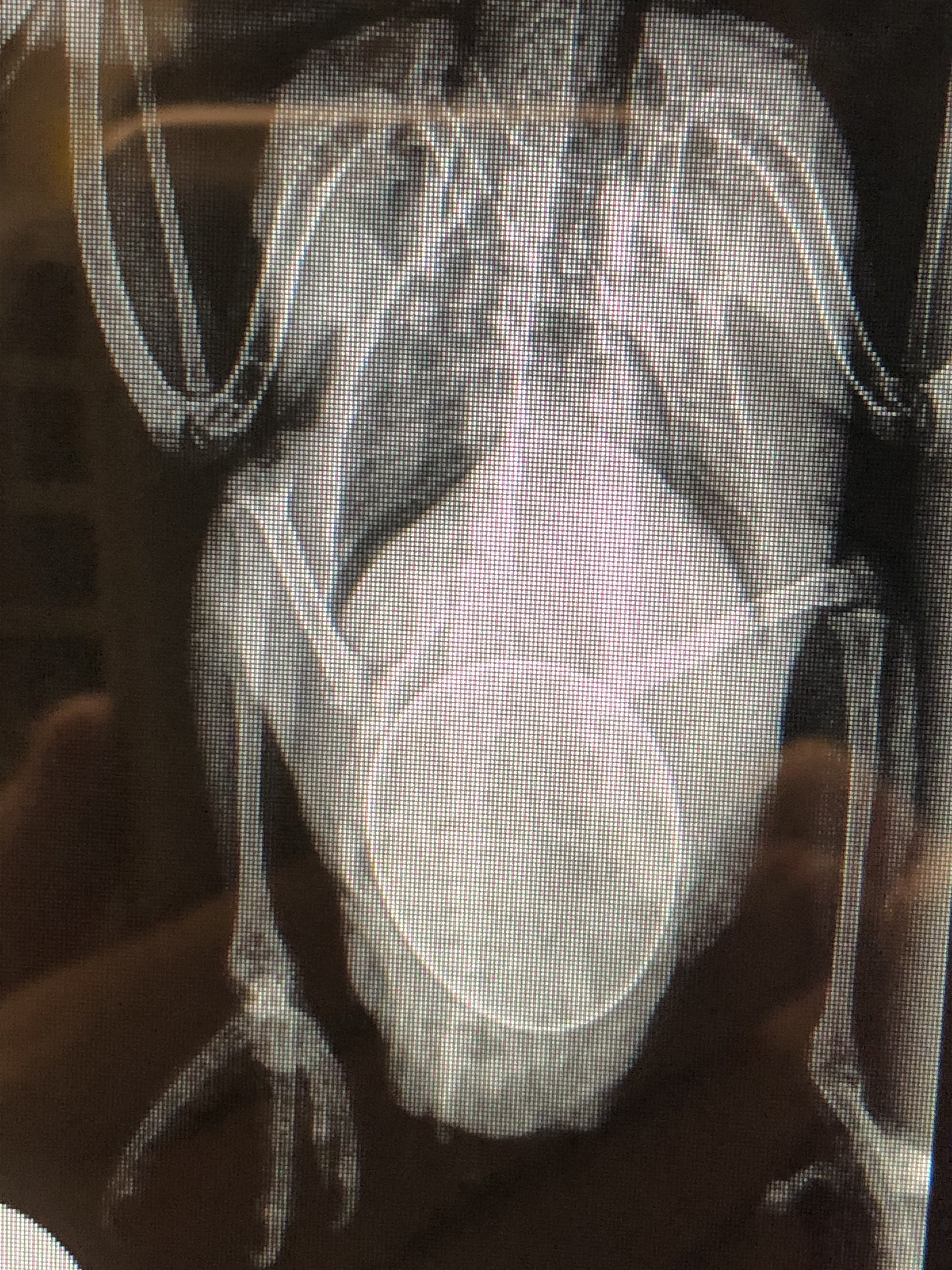
You see, the Dalmatian breed of dog has a unique inherited predisposition to uric acid bladder stones. Foods like organ meat or seafood are high in a form of protein called purines. The liver is supposed to break this protein down into a harmless substance that the body absorbs but in Dalmatians, lacking this gene, the liver only processes the protein into uric acid, which is filtered through the kidneys and settles out as small stones (as hard as coarse sand) into the bladder. The best way I can draw this picture with words is to compare the Dalmatian, a mammal, to birds. Birds are uricotelic, meaning they do not break down body waste into urine but instead their kidneys excrete uric acid, the solid white matter you see in a bird dropping. This is normal for a bird. But mammals – dogs – are supposed to excrete waste filtered through the kidneys as a thin fluid, urine. Dalmatian dogs make urine, but some will also make uric acid crystals which build up in the kidneys and bladder. Hundreds of these crystals can be deposited inside the bladder and obstruct the male Dalmatian dog’s ability to urinate. Once the bladder outlet is obstructed the dog can’t urinate and the bladder will slowly fill as the kidneys continue to produce both urine and uric acid. Without relief of the obstruction the bladder will rupture and/or the kidneys shut down, both potentially leading to death.

Spotty’s abdominal x-ray showed an enormous bladder, though the uric acid stones causing the problem weren’t well defined. His bloodwork demonstrated kidney distress, but within parameters that we felt could be reversed to normal once his obstruction was surgically cleared. We whisked Spotty in for emergency surgery, relieving his obstruction and emptying his bladder of hundreds of tiny grey stones. Intravenous fluids helped get his kidneys up and running again, a process we call diuresis. The stones or uroliths were analyzed to be certain they were uric acid in nature and a proper prevention plan put in place.

We know that high protein, meat-based diets may not be the best choice for any Dalmatian at this time as many are formulated with high purine foods of organ meat or seafood. Avoiding these purine-rich foods puts less burden on the liver and can greatly decrease the formation of stones. Dietary avoidance of these foods is a cornerstone to prevention.

This condition is similar to the disease of gout in people. In people the uric acid crystals settle in the joints, causing inflammation and pain. Interestingly, the same medication may be used to treat both humans and canines.

Spotty is lucky. Through a low-protein, non-meat based diet, medication and monitoring of his urine he is expected to lead a full, normal life. Because the gene has been identified, Dalmatian breeders can now specifically test and work to breed out this genetic mutation. The Veterinary Genetics Lab at the University of California, Davis, School of Veterinary Medicine offers genetic testing for a variety of breed-inherited diseases.

Christine B. McFadden, DVM
drmc@mcmenagerie.com


Ear Jam / Otitis Externa

Today we will tackle the shaky subject of ear infections in the dog. Ear infections rank right up there with some of the smelliest problems you might ever endure with your pet. And your dog feels a lot worse about it. The ear is exquisitely built in the dog – those long, silky flaps (pinnae) framing your dog’s face, so pettable, tilting to make clown faces, waving in the wind during a glorious ride peeking out the car window. Some ears stand upright at attention and a few are bald. All ears are beautiful. The inner workings of the ear contain the mysteries that lead to one of the major 5 senses in the body : Hearing. A dogs hearing ability is considered to be 4 times that of the average human being. Sound is caused by air vibrations – the more vibrations per second, the higher the sound. Humans are limited to hearing sounds that vibrate at 20,000 vibrations per second, while dogs pick them up at 50,000 vibrations per second. Another factor is that people have only 8 ear muscles to help wiggle their ears to funnel sound in and capture noises ; the dog has 18 muscles (Not to be outdone, the cat, with 30 ear muscles, enjoys the best hearing of all!).

Did you know that dogs lack most of the skin glands that a person has? One place that they DO have true skin glands is lining the inner ear canal or passageway into the ear. If you gently lift your pets ear flap, or pinnae, you’ll see a hole at the base which goes into the head: this is the ear canal. The average ear canal is 2 inches long! In the dog it travels down, with a little L-shaped jog at the end just above the ear drum (tympanic membrane). This may serve to protect the ear drum from things that fall into the ear. Just as in a person, the ear drum is exquisitely sensitive, a barrier between the outer ear and the middle and inner ear where the hearing organs are located. This fine membrane vibrates in response to the sound waves it receives, transmitting them to the middle and inner ear for complex translation. Any fluid, foreign object, mass, or swelling inside the ear may deflect the passage of air vibrations and alter hearing. Long term infections may lead to permanent thickening (hyperplasia) of the canal, blocking the travel of sound.  Loss of elasticity to the ear drum may result in deafness to varying degrees. Ear infections are serious.

So what can go wrong with an ear? How can you tell? One of the easiest things to remember is that dogs don’t really make ear wax like people do – so if you see yellow or black discharge lining the ear you know something is very wrong. This ceruminous or glandular discharge may be made up of several bacteria or yeast or both. Some people will note a bread-like odor if yeast is present, and one bacteria, pseudomonas, is quaintly referred to by its “rotten taco smell”. Because the average length of a dog’s ear canal is over 2 inches long, a veterinarian requires a specialized instrument, the otoscope, to examine deeply. An otoscope has a strong halide light and magnifying lens, coupled with a long tube sized for the individual to look deep into the canal for the cause of the problem.

Like something out of a cheap horror movie, sometimes we put our eyeball to the scope and see giant (magnified) grey legs waving at us – ear mites, Otobius megnini. These are not to be confused with the cat ear mite, Otodectes cynotis, which would be about the size of a small grain of pepper if visible at all and never waves at you. We don’t see them that often around Merced and it’s usually good for a surprised shriek. In the USA the Otobius mite is most common in the arid regions of the Southwest and has a preference for the ear, even, occasionally, in people.

Very commonly we find foxtails buried deep into ears, painfully jabbing the tender canal. Sometimes they can be removed with a long narrow instrument called an “alligator forceps” – your pet will let you know if sedation is required! The pointed, piercing end of a foxtail awn must feel like an ice pick is poking into their head! Foxtails in a deep ear hole are never good, as they may perforate the ear drum and travel into the head, adjacent jaw or lymph nodes. The ear responds vigorously to this foreign body, mounting a massive infection in an attempt to eject the invader; alas, to no avail as gravity keeps it buried deep in the canal. The best news about foxtails is that once removed the infections are usually responsive to medication and clear up quickly.

Chronic, recurring ear infections are extremely common in our Valley. Properly called “otitis externa”, these infections bear no relationship to the inner ear infections young children often suffer from. They do not respond to oral antibiotics given to toddlers. They are not similar infections at all. Hayfever, or Atopy, may be behind much of the recurring or chronic infections of dogs. The ear canal of a dog is lined with true glands. Glandular tissue may respond to an allergen, like pollens in the air (and is our Valley not King of Pollen?) by becoming inflamed. Some breeds, like the Labrador Retriever and Cocker Spaniel, are genetically born with twice as many glandular cells lining their ear canals as other breeds, making them even more susceptible to ear infections. When the ear canal is examined we can see the tiny blood vessels swollen and red and the little glands puff up, making the inner ear canal look bumpy like a cobblestone road. As the tissue swells, a thin fluid exudate oozes out, creating a warm, wet environment deep in the ear hole – perfect for breeding yeast and bacteria. And do they ever! This ear soup is stuck, since it can’t drain by itself. If unattended, blood and pus may form. These infections are very painful, and a dog may paw or shake its head so hard the blood vessels burst and the pinnae fills up like a balloon with a blood hematoma. Polyps and cancerous masses can occur. All ear infections require careful treatment plans only your veterinarian can prescribe – your pets smiling face and happy head wag is its own reward!

Christine B. McFadden, DVM

drmc@mcmenagerie.com

Cough, Hack, Gag, Enough!


Dr Mc Blog

October 4, 2014

Today we are referencing the coughing dog. Dogs only; cats, which are indeed a world unto themselves, need not apply. Cats want their own column. Besides, there’s lots more coughing dogs out there and plenty to discuss about each of the many possibilities behind a dog’s cough.

First, it can be surprisingly hard to determine if the complaint IS a true cough. A cough should sound like an outward sort-of-explosive breath. If smokers hadn’t been virtually banned from California, everyone would know what a true cough sounds like, but as it is there is room for doubt.  Always remember that dogs have no social compulsion to be dainty and will not try to subvert a cough into a fake sneeze or whistle. They “hack”. They may gag, but here we enter a grey area, as a dog that can’t draw breath well enough may almost vomit – but most of the time chest symptoms are different than stomach issues. I am filled with admiration for the client who can graphically imitate their pet’s cough in the clinic. On occasion I defer to the ubiquitous cell phone and request the client record a home video of the act. Is the cough productive (think wet, slimey goop coming out, maybe colored yellow or with blood) or dry?

Once we have established that a true cough is present, we can mull over the algorithm or medical pathway that leads to a diagnosis. Internal medicine is like a treasure hunt. If you enjoyed “CandyLand” as a child you will love going from clue (symptoms) to clue (diagnostic tests) as you follow the path to final diagnosis and treatment.

There is the heart cough, when leaky valves inside the heart don’t regulate blood flow properly and fluid pools in the lungs. The lungs, unsuccessfully, try to cough the excess fluid out. Or the heartworm cough. The heartworm, Dirofilaria immitis, is a mosquito transmitted worm that grows in the bloodstream and nests in the heart and lungs. Signs frequently include shortness of breath and coughing.

Bronchitis occurs in dogs, both allergic or infectious : viral, bacterial, fungal. The infamous diagnosis “Kennel Cough” springs from this group. Valley Fever falls into the fungal group. True bacterial pneumonia is relatively rare in dogs.

Foreign objects that have been aspirated or breathed into the chest, like our Valley foxtail weed awn, can set up an infection that may lead to coughing.

Chest masses and cancers, whether primary from the lung itself or metastasized, having spread to the lungs from another cancer site in the body, will take up physical space in the chest and lead to a cough.

Less malign causes are sometimes as simple as breed/body type predisposing conditions. Some breeds have a hypoplastic, very tiny trachea – it’s like trying to breathe through a straw. Others may suffer from a collapsing trachea. These conditions are common in both tiny and brachygnathic (pushed-in face) breeds – think Yorkies and Olde English Bulldogs.

I’ll tell you Jade’s story. Jade is a bouncy tricolor Rat Terrier who rarely kept more than 3 feet on the ground as she danced around. That day in February she was unusually quiet. In addition to that, her owner had heard a soft cough at home. I heard muffled lung sounds when I listened through my stethoscope. Hmmm, no leaky  heart valves and she’d had a negative heartworm test a few months back before starting on preventative medicine. We took chest x-rays, which showed a cloudy area on one side of her chest. Her bloodwork showed no sign of infection or anemia. I asked all the usual questions, but was stumped. Permission to do a chest tap to look at the fluid was granted. We drew out pure blood! Despite the negative answer to questions about mouse bait exposure, we tested Jade to see if her blood was clotting – and it wasn’t! I diagnosed a rodenticide toxicity. The missus had been doing some spring cleaning two days earlier and found an empty box of mouse bait in an unused closet. She’d tossed it without another thought.  Many mouse baits stop the body’s ability to clot blood and the animal that eats it bleeds to death, ANY animal. Bleeding may start anywhere, though it was unusual that Jade began to bleed only into her chest. The good news is that all veterinary clinics keep the antidote on hand and within hours Jade was making a full recovery. Her cough disappeared as quickly as it came.

If your dog is coughing Jade strongly recommends that you consult your veterinarian early on. We have only touched on a few of the diseases that share the symptom “coughing”. Stakes can be very high with a dog that coughs. Had her owner waited, Jade would not have been able to share her story with you today.

Christine B. McFadden, DVM
Valley Animal Hospital, Merced